Copyright
©The Author(s) 2024.
World J Clin Cases. Nov 6, 2024; 12(31): 6479-6485
Published online Nov 6, 2024. doi: 10.12998/wjcc.v12.i31.6479
Published online Nov 6, 2024. doi: 10.12998/wjcc.v12.i31.6479
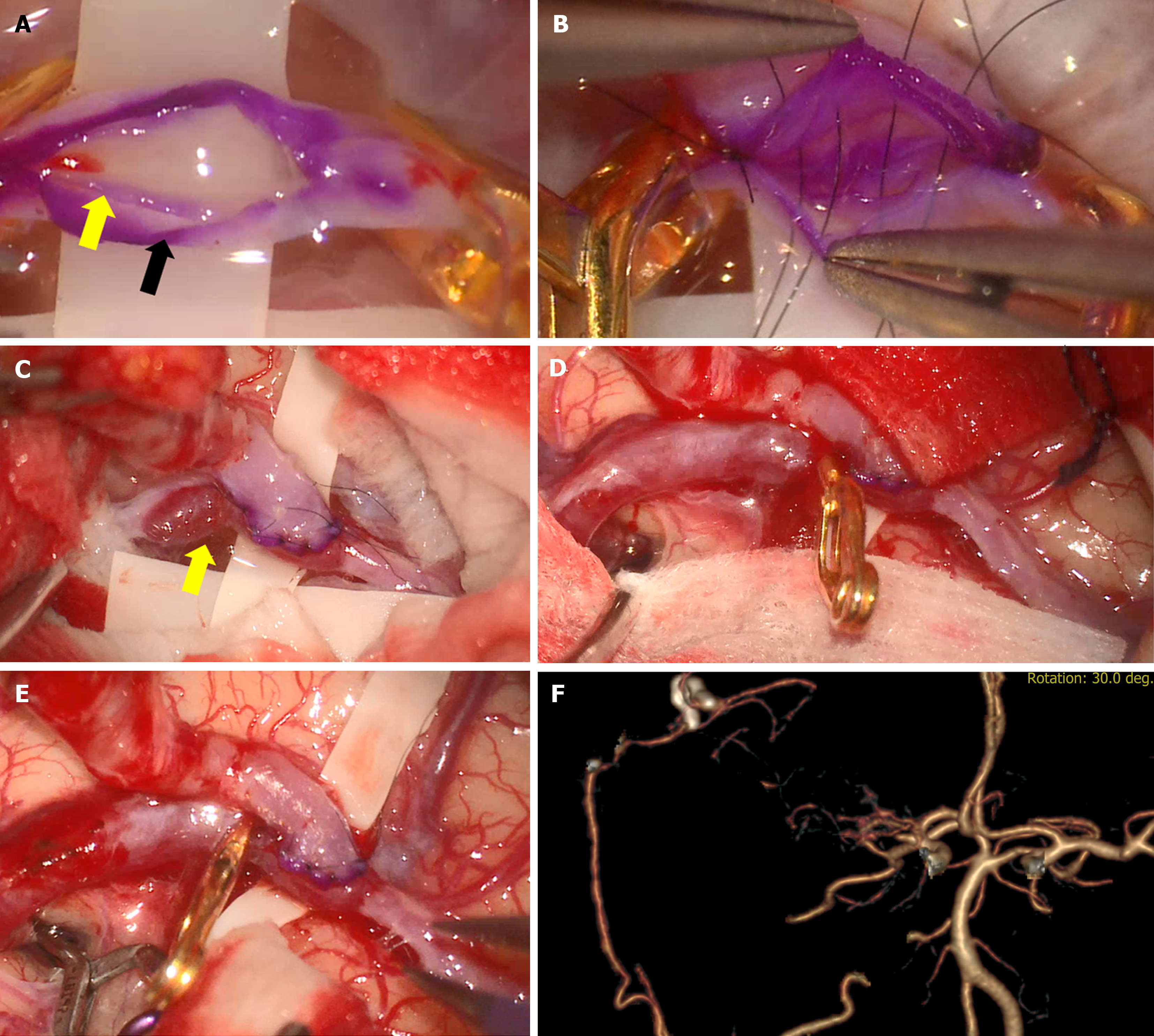
Figure 2 Intra- and postoperative image findings (case 1).
A: Intima and media (yellow arrow) of recipient artery dissected from the adventitia layer (black arrow) after arteriotomy; B: To prevent dissection, we performed immediate suturing to ensure that the intima and adventitia were not separated; C: Dissecting aneurysm (yellow arrow) occurred at the anastomosis site after temporary clip removal at the recipient artery; D: Pseudoaneurysm gradually progressed; E: Before further dissection, the dissected portion was trapped using a clip; F: Postoperative brain computed tomography angiography confirmed arterial anastomosis between superficial temporal artery and distal middle cerebral artery.
- Citation: Lee YJ, Park W, Joo SP. Recipient artery dissection during extracranial-intracranial bypass surgery: Two case reports. World J Clin Cases 2024; 12(31): 6479-6485
- URL: https://www.wjgnet.com/2307-8960/full/v12/i31/6479.htm
- DOI: https://dx.doi.org/10.12998/wjcc.v12.i31.6479


