Copyright
©The Author(s) 2024.
World J Clin Cases. Nov 6, 2024; 12(31): 6479-6485
Published online Nov 6, 2024. doi: 10.12998/wjcc.v12.i31.6479
Published online Nov 6, 2024. doi: 10.12998/wjcc.v12.i31.6479
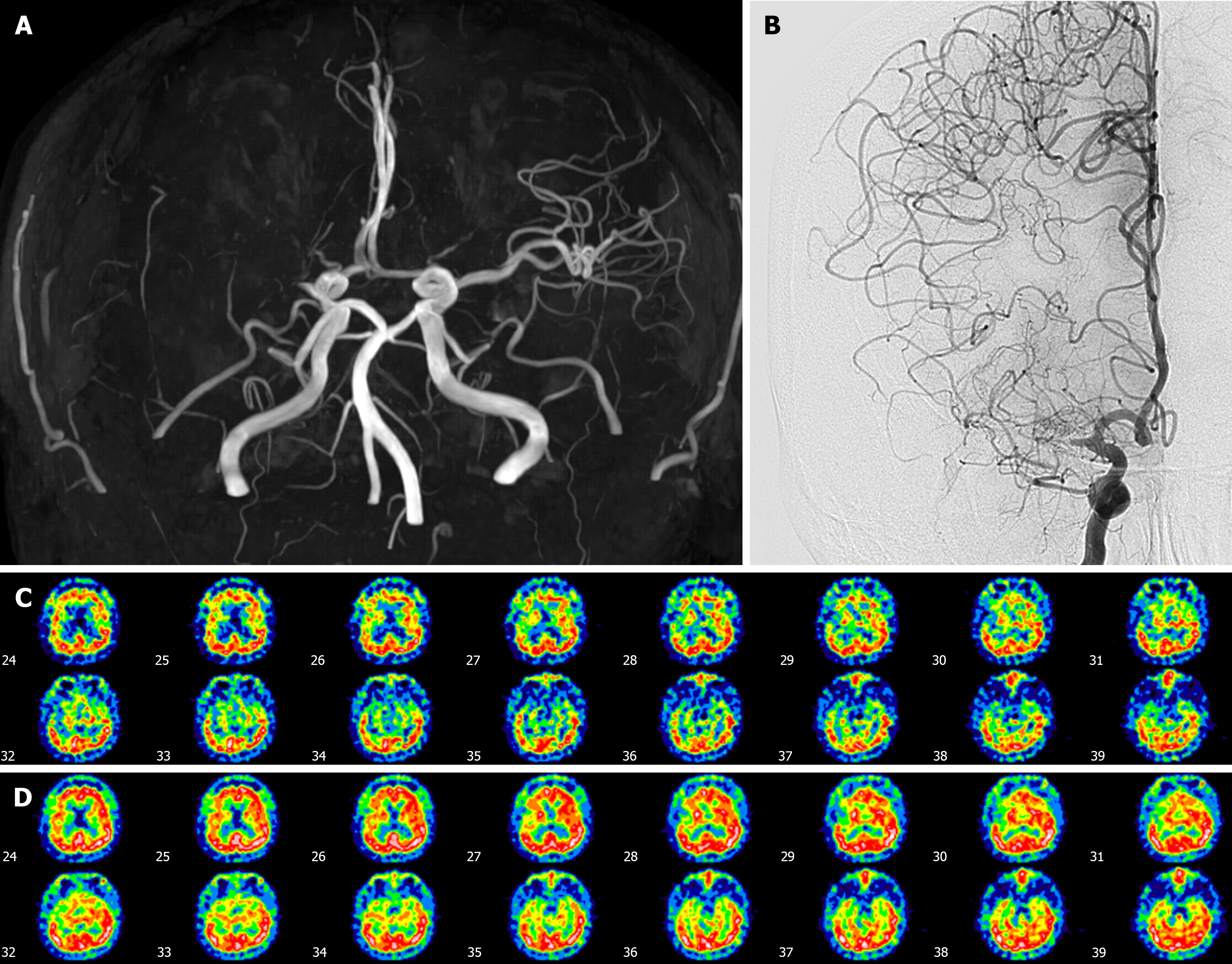
Figure 1 Preoperative brain image findings (case 1).
A: Initial brain magnetic resonance angiography revealing right middle cerebral artery occlusion; B: Diagnostic cerebral angiography of the right internal carotid artery injection demonstrating proximal right middle cerebral artery occlusion and no transdural collaterals; C: Brain single photon emission computed tomography revealing moderately diminished baseline perfusion in the right frontal lobe; D: Moderately diminished cerebrovascular reserve in the right frontal, parietal, and temporal lobes.
- Citation: Lee YJ, Park W, Joo SP. Recipient artery dissection during extracranial-intracranial bypass surgery: Two case reports. World J Clin Cases 2024; 12(31): 6479-6485
- URL: https://www.wjgnet.com/2307-8960/full/v12/i31/6479.htm
- DOI: https://dx.doi.org/10.12998/wjcc.v12.i31.6479


