Copyright
©The Author(s) 2024.
World J Clin Cases. Nov 6, 2024; 12(31): 6462-6471
Published online Nov 6, 2024. doi: 10.12998/wjcc.v12.i31.6462
Published online Nov 6, 2024. doi: 10.12998/wjcc.v12.i31.6462
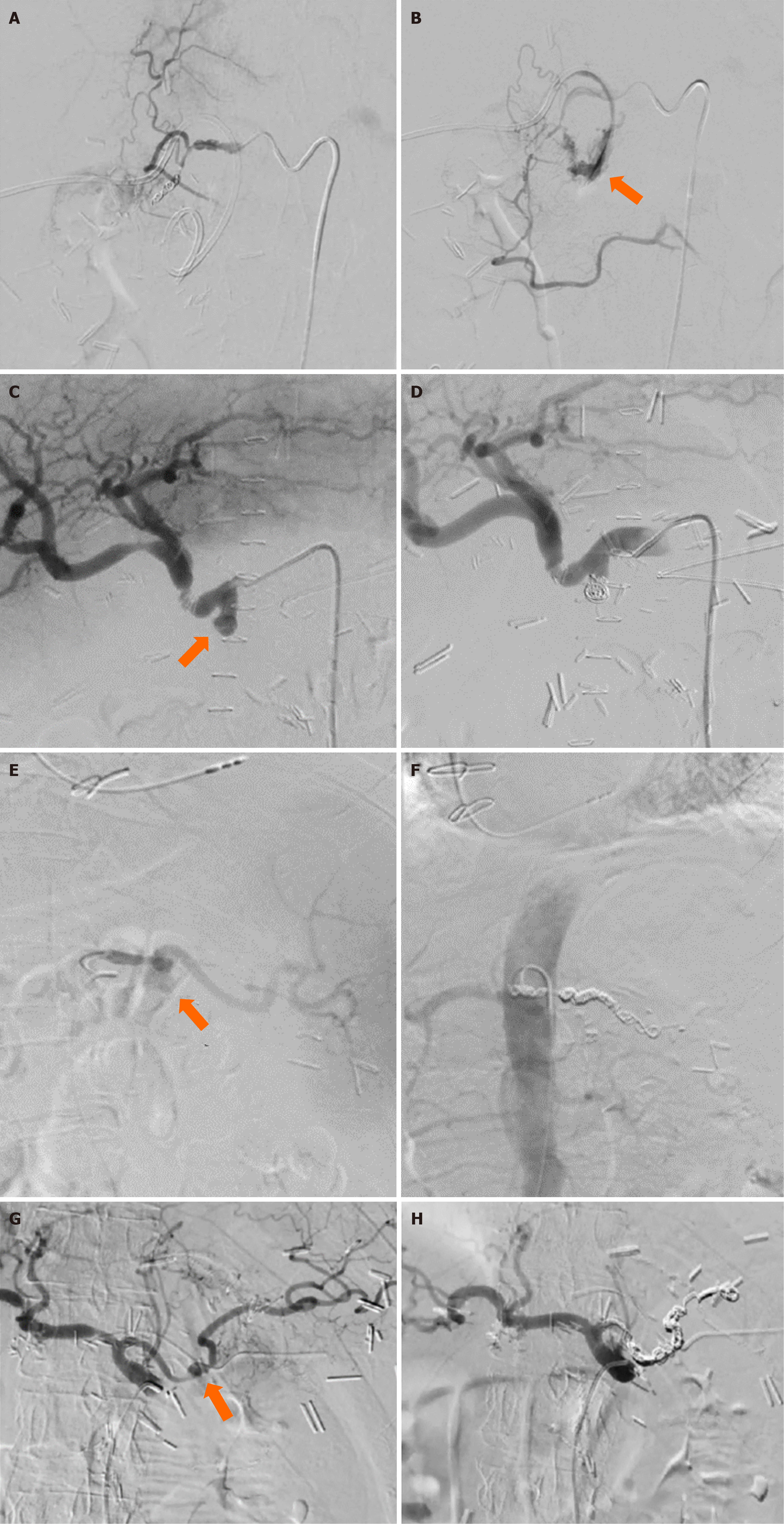
Figure 2 Digital subtraction angiography.
A: Digital subtraction angiography shows extravasation of the intravenous contrast from the gastroduodenal artery (arrow); B: Successful obliteration of the gastroduodenal artery stump with endovascular coiling; C: 7 mm visceral arterial pseudoaneurysm of the gastroduodenal artery stump (arrow); D: Successful embolization of the gastroduodenal artery pseudoaneurysm using endovascular coiling; E: Visceral arterial pseudoaneurysm of the splenic artery (arrow); F: Successful obliteration of the splenic artery using endovascular coils; G: Digital subtraction angiography shows 18 mm visceral arterial pseudoaneurysm of the splenic artery (arrow); H: Successful obliteration of the splenic artery using endovascular co.
- Citation: Petrovic I, Romic I, Alduk AM, Ticinovic N, Koltay OM, Brekalo K, Bogut A. Delayed postpancreatectomy hemorrhage as the role of endovascular approach: Four case reports. World J Clin Cases 2024; 12(31): 6462-6471
- URL: https://www.wjgnet.com/2307-8960/full/v12/i31/6462.htm
- DOI: https://dx.doi.org/10.12998/wjcc.v12.i31.6462


