Copyright
©The Author(s) 2024.
World J Clin Cases. Jan 26, 2024; 12(3): 665-670
Published online Jan 26, 2024. doi: 10.12998/wjcc.v12.i3.665
Published online Jan 26, 2024. doi: 10.12998/wjcc.v12.i3.665
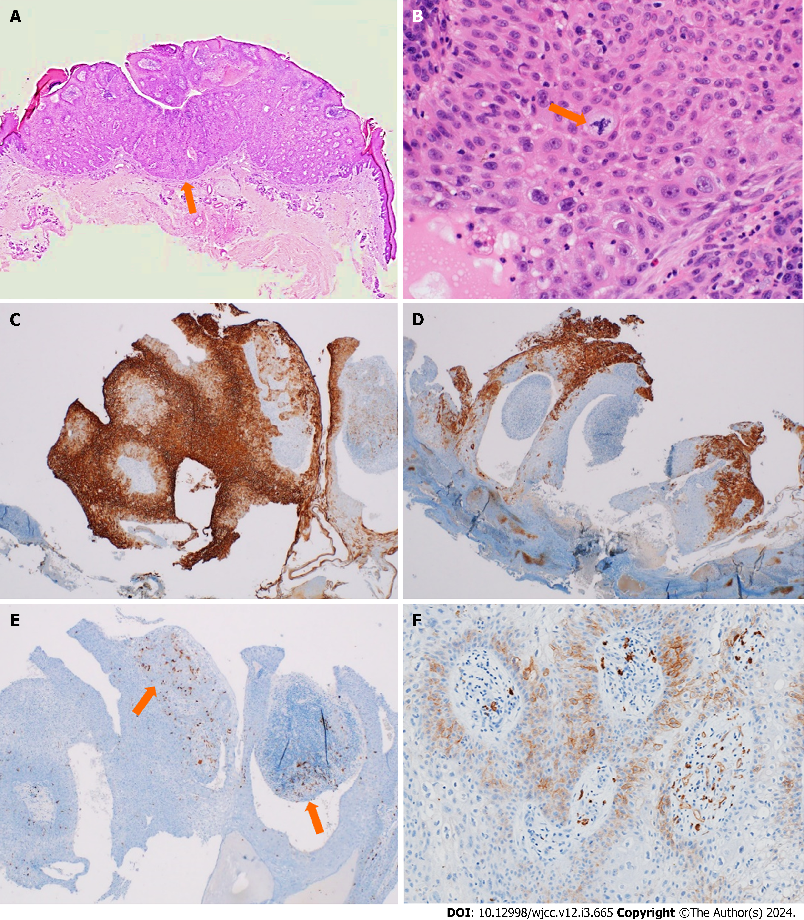
Figure 4 Histological features of porocarcinoma.
A: Epidermal tumor cluster arising from the intraepidermal portion of the sweat gland ducts and extending to the dermal portion (arrow); B: The tumor cells exhibit atypical mitosis (arrow); C-F: Tumor cells are positive for epithelial membrane antigen (C), cytokeratin 7 (D), focal expression for S-100 (E) on scattered melanocytes (arrows), and C-kit (F). A and B: Hematoxylin and eosin; C-F: Immunohistochemical staining of porocarcinoma. Original magnification: A (× 20); B (× 400); C-E (× 40); F (× 100).
- Citation: Lim SB, Kwon KY, Kim H, Lim SY, Koh IC. Porocarcinoma in a palm reconstructed with a full thickness skin graft: A case report. World J Clin Cases 2024; 12(3): 665-670
- URL: https://www.wjgnet.com/2307-8960/full/v12/i3/665.htm
- DOI: https://dx.doi.org/10.12998/wjcc.v12.i3.665


