Copyright
©The Author(s) 2024.
World J Clin Cases. Jan 26, 2024; 12(3): 616-622
Published online Jan 26, 2024. doi: 10.12998/wjcc.v12.i3.616
Published online Jan 26, 2024. doi: 10.12998/wjcc.v12.i3.616
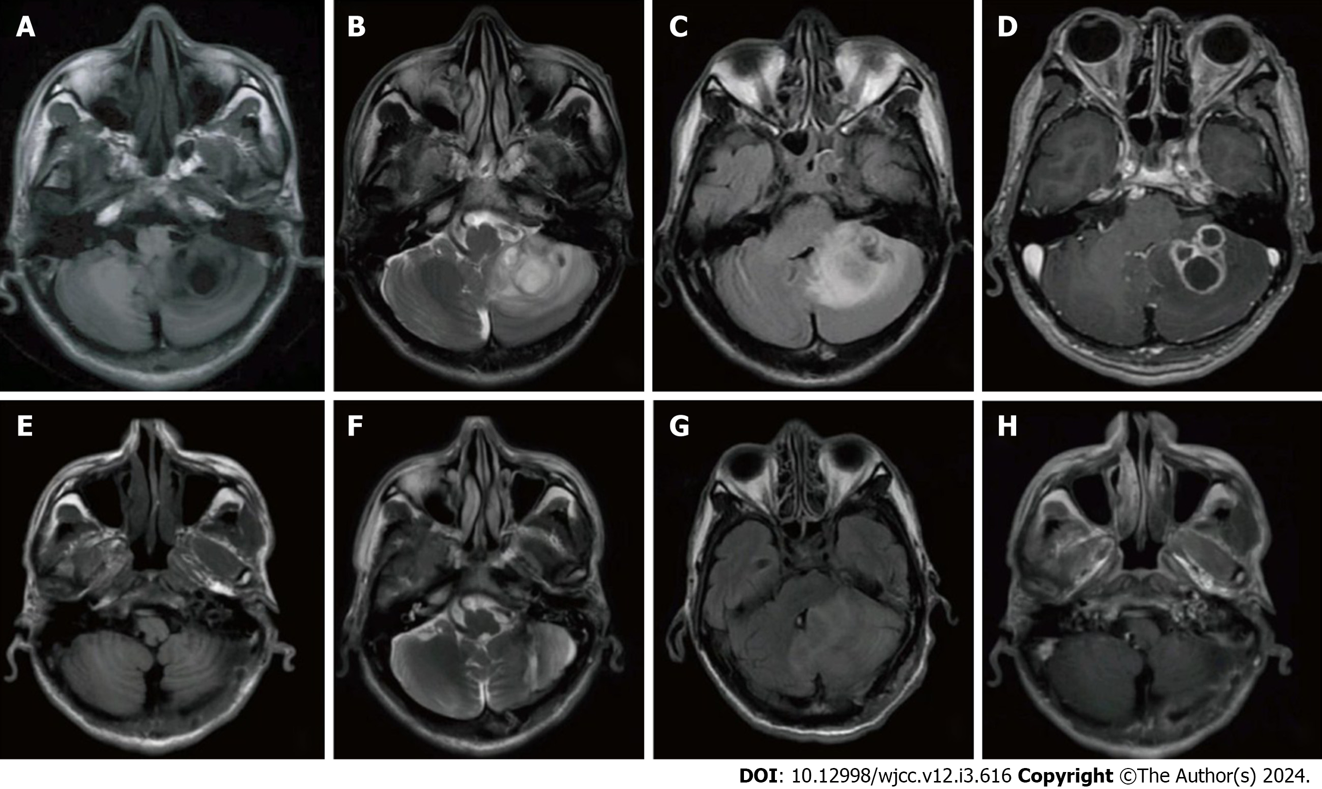
Figure 1 The patient's brain magnetic resonance imaging results before treatment and follow-up.
A and E: Long T1-weighted imaging signal in left cerebellar hemisphere before treatment; B and F: Long T2-weighted imaging signal in left cerebellar hemisphere in follow-up; C: High T2-fluid-attenuated inversion recovery signal in left cerebellar hemisphere before treatment; D: Enhancement showing a multiventricular ring-like enhancement in left cerebellar hemisphere before treatment; G: Slightly high T2-FLAIR signal in left cerebellar hemisphere in follow-up; H: Enhancement showing a ring enhancement in left cerebellar hemisphere in follow-up. MRI: Magnetic resonance imaging; T1WI: T1-weighted imaging; T2WI: T2-weighted imaging; FLAIR: Fluid-attenuated inversion recovery.
- Citation: Zhu XM, Dong CX, Xie L, Liu HX, Hu HQ. Brain abscess from oral microbiota approached by metagenomic next-generation sequencing: A case report and review of literature. World J Clin Cases 2024; 12(3): 616-622
- URL: https://www.wjgnet.com/2307-8960/full/v12/i3/616.htm
- DOI: https://dx.doi.org/10.12998/wjcc.v12.i3.616


