Copyright
©The Author(s) 2024.
World J Clin Cases. Jan 26, 2024; 12(3): 607-615
Published online Jan 26, 2024. doi: 10.12998/wjcc.v12.i3.607
Published online Jan 26, 2024. doi: 10.12998/wjcc.v12.i3.607
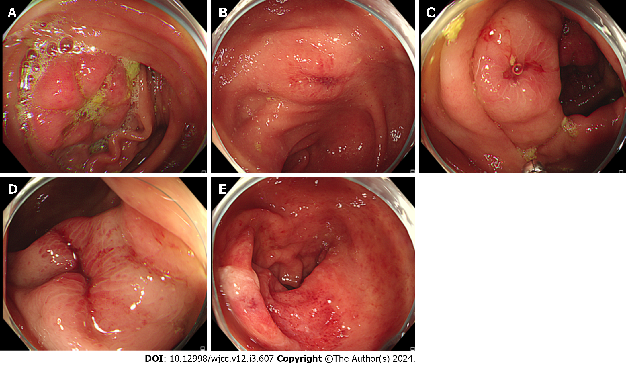
Figure 1 Shows the colonoscopy of 2021-06-28.
A: Flaky mucosal hyperemia and edema were observed in ileocecal region, accompanied by ulcer formation; B: The ascending colon is scattered with ulcers and surrounding mucous membrane flaky hyperemia and edema; C: The ascending colon is scattered with ulcers and surrounding mucous membrane flaky hyperemia and edema; D: A 3 cm long longitudinal ulcer was observed 65 cm from the anal margin, and the surrounding mucosa was hyperemia, edema and erosion; E: Rectum mucosa hyperemia edema, vascular texture blurred, multiple erosion and shallow ulcer formation.
- Citation: Wang CL, Si ZK, Liu GH, Chen C, Zhao H, Li L. Ischemic colitis induced by a platelet-raising capsule: A case report. World J Clin Cases 2024; 12(3): 607-615
- URL: https://www.wjgnet.com/2307-8960/full/v12/i3/607.htm
- DOI: https://dx.doi.org/10.12998/wjcc.v12.i3.607


