Copyright
©The Author(s) 2024.
World J Clin Cases. Jan 26, 2024; 12(3): 565-574
Published online Jan 26, 2024. doi: 10.12998/wjcc.v12.i3.565
Published online Jan 26, 2024. doi: 10.12998/wjcc.v12.i3.565
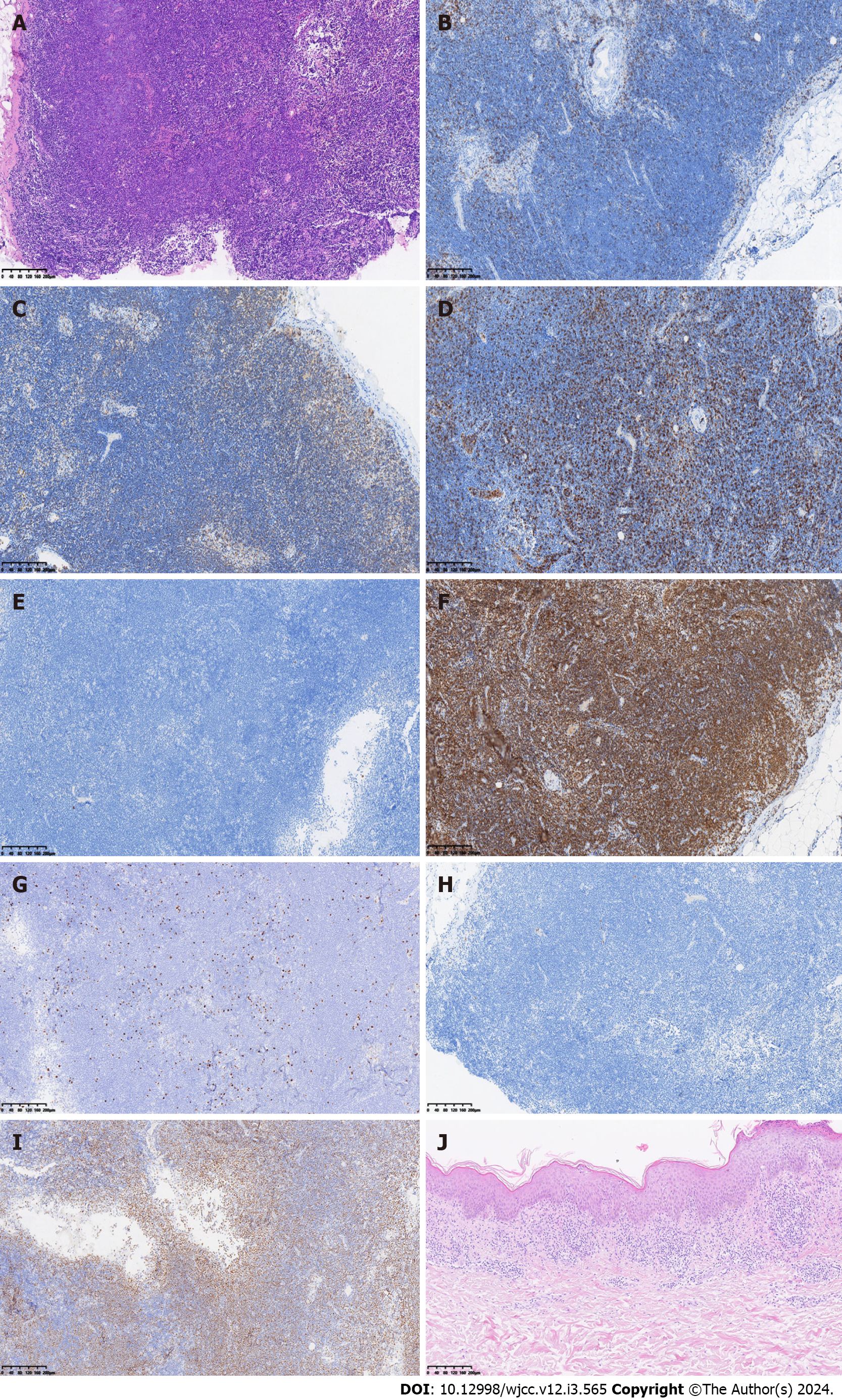
Figure 3 Immunohistochemical staining of pathological biopsy at diagnosis.
A: Hematoxylin-eosin (HE) staining of lymph nodes at 10 × magnification; B: CD3 (partial +); C: CD20 (+); D: CD5 (partial +); E: CD23 (-); F: Bcl-2 (+); G: Ki67 (about 15%); H: CyclinD1 (-); I: P53 (about 60%); J: HE staining of the skin at 10 × magnification. Immunohistochemical staining of lymph nodes at 10 × magnification (B-I).
- Citation: Bai SJ, Geng Y, Gao YN, Zhang CX, Mi Q, Zhang C, Yang JL, He SJ, Yan ZY, He JX. Marginal zone lymphoma with severe rashes: A case report. World J Clin Cases 2024; 12(3): 565-574
- URL: https://www.wjgnet.com/2307-8960/full/v12/i3/565.htm
- DOI: https://dx.doi.org/10.12998/wjcc.v12.i3.565


