Copyright
©The Author(s) 2024.
World J Clin Cases. Jan 26, 2024; 12(3): 551-559
Published online Jan 26, 2024. doi: 10.12998/wjcc.v12.i3.551
Published online Jan 26, 2024. doi: 10.12998/wjcc.v12.i3.551
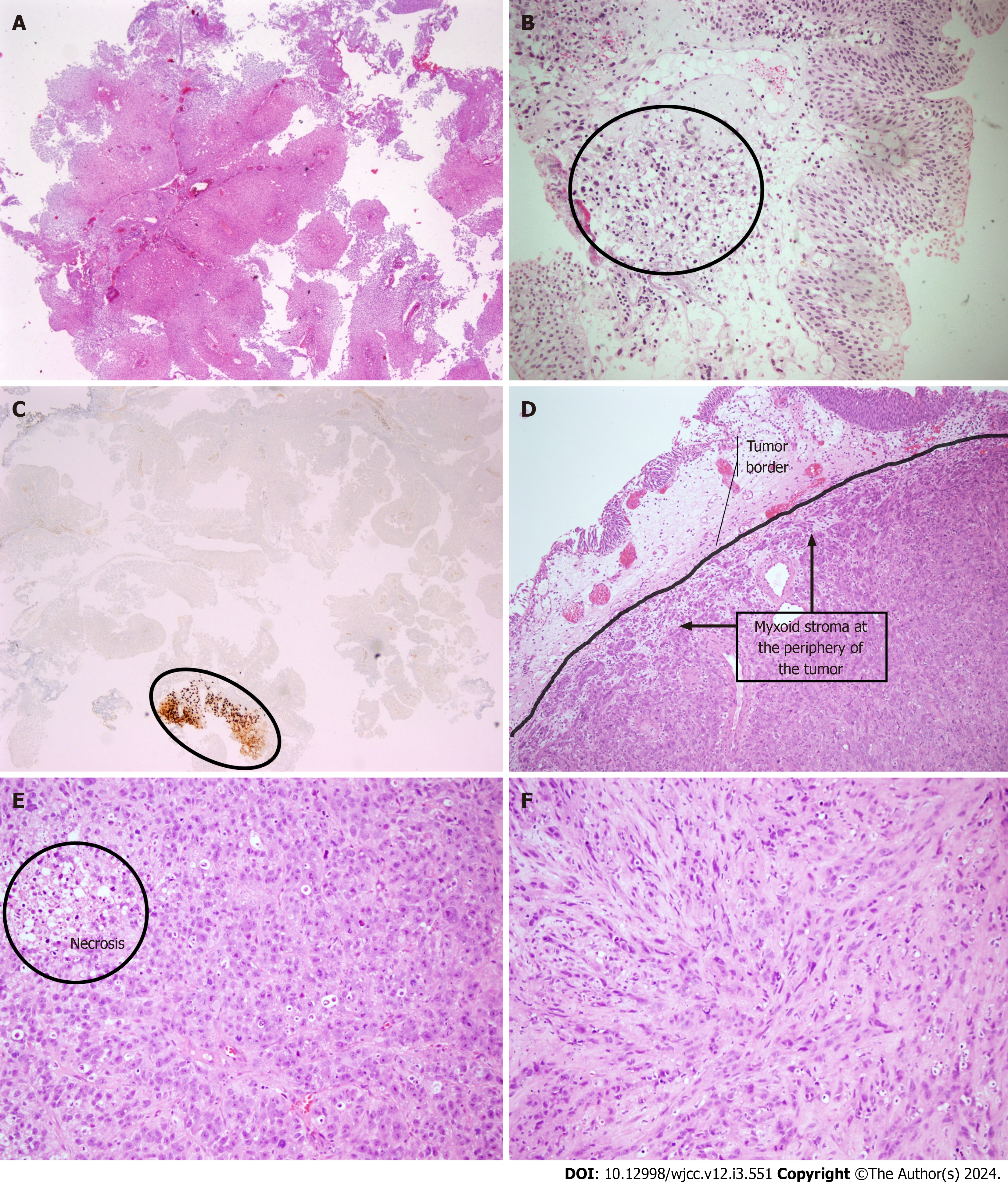
Figure 1 Epithelioid malignant peripheral nerve sheath tumor of the bladder.
A: Non-invasive low grade papillary urothelial carcinoma seen on initial biopsy; B: Non-invasive low grade papillary urothelial carcinoma and atypical mesenchymal cell infiltration in the lamina propria determined on second biopsy (the area within the circle); C: SOX-10 immunopositivity in atypical mesenchymal cells (the area annotated with ellipse) located in the lamina propria in one of the tissue fragments representing the non-invasive papillary urothelial carcinoma; D: Focal myxoid stroma at the periphery of the tumor with a lobular growth pattern (an arc is used to distinguish normal urothelium from tumoral tissue); E: Epithelioid tumor cells with round vesicular nuclei, prominent nucleoli and abundant eosinophilic cytoplasm; necrosis in the upper left corner; F: Spindle tumor cells with fascicular growth (hematoxylin and eosin, SOX10, × 40, × 100 and × 200).
- Citation: Ozden SB, Simsekoglu MF, Sertbudak I, Demirdag C, Gurses I. Epithelioid malignant peripheral nerve sheath tumor of the bladder and concomitant urothelial carcinoma: A case report. World J Clin Cases 2024; 12(3): 551-559
- URL: https://www.wjgnet.com/2307-8960/full/v12/i3/551.htm
- DOI: https://dx.doi.org/10.12998/wjcc.v12.i3.551


