Copyright
©The Author(s) 2024.
World J Clin Cases. Oct 16, 2024; 12(29): 6327-6334
Published online Oct 16, 2024. doi: 10.12998/wjcc.v12.i29.6327
Published online Oct 16, 2024. doi: 10.12998/wjcc.v12.i29.6327
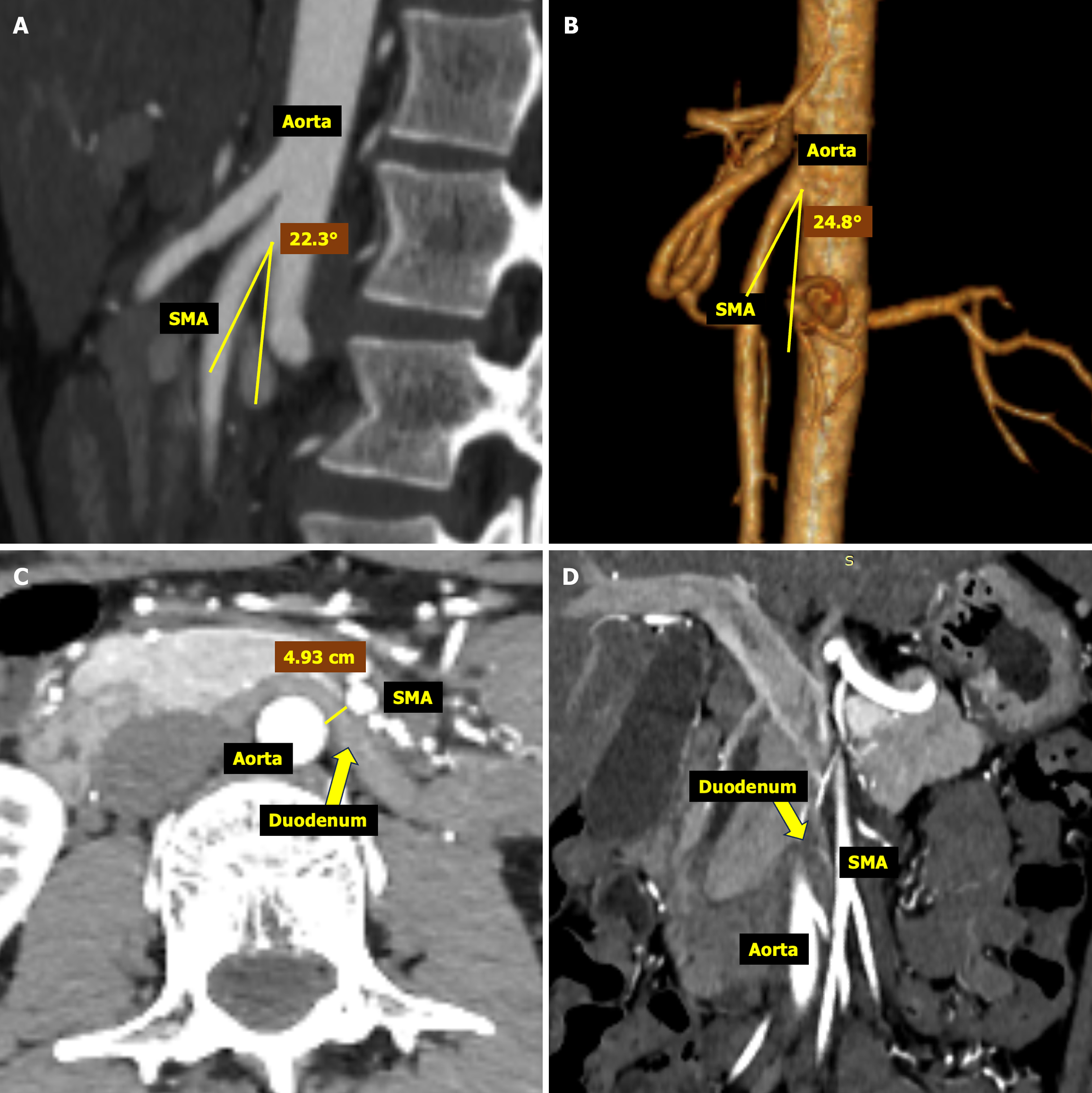
Figure 2 Follow-up abdominal computed tomography angiography examination.
A-D: Computed tomography angiography image (A) and the three-dimensional reconstruction of abdominal artery (B) showed the angle between the superior mesenteric artery (SMA) and the aorta; Axial (C) and coronal (D) images illustrate the distance between the horizontal plane of the aorta and SMA at the level of the duodenum. SMA: Superior mesenteric artery.
- Citation: Liu B, Sun H, Liu Y, Yuan ML, Zhu HR, Zhang W. Comprehensive interventions for adult cyclic vomiting syndrome complicated by superior mesenteric artery syndrome: A case report. World J Clin Cases 2024; 12(29): 6327-6334
- URL: https://www.wjgnet.com/2307-8960/full/v12/i29/6327.htm
- DOI: https://dx.doi.org/10.12998/wjcc.v12.i29.6327


