Copyright
©The Author(s) 2024.
World J Clin Cases. Oct 16, 2024; 12(29): 6320-6326
Published online Oct 16, 2024. doi: 10.12998/wjcc.v12.i29.6320
Published online Oct 16, 2024. doi: 10.12998/wjcc.v12.i29.6320
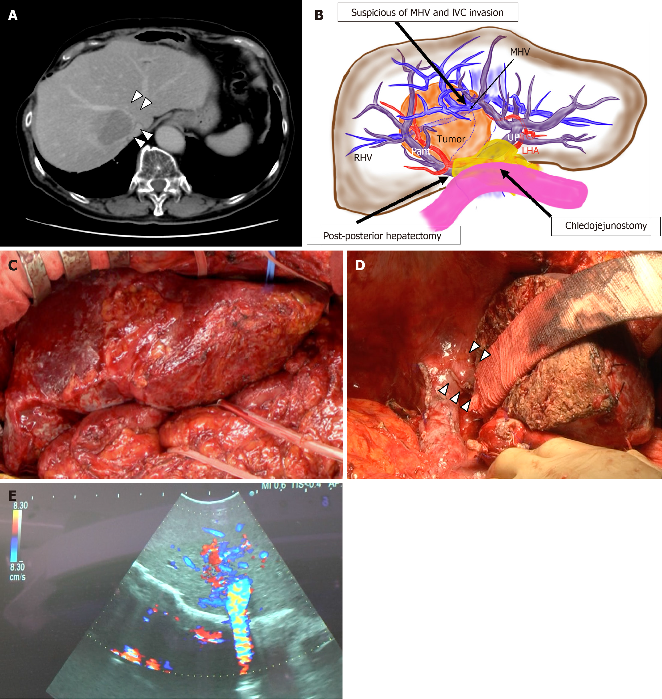
Figure 1 Preoperative and the first operation information of this case.
A: Contrast enhanced computed tomography of tumor. Arrowhead indicates suspicious of tumor invasion for middle hepatic vein and inferior vena cava; B: Schema of this case; C: Intraoperative photography after adhesiolysis around the liver; D: Intraoperative photography after specimen is removed. Arrowhead indicates extrahepatic naked left hepatic vein; E: Doppler ultrasonography shows normal hepatic vein flow direction. MHV: Middle hepatic vein; IVC: Inferior vena cava; RHV: Right hepatic vein; UP: Umbilical portion, LHA: Left hepatic artery; Pant: Anterior branch of portal vein.
- Citation: Higashi H, Abe Y, Abe K, Nakano Y, Tanaka M, Hori S, Hasegawa Y, Yagi H, Kitago M, Kitagawa Y. Novel procedure for hepatic venous outflow block after liver resection: A case report. World J Clin Cases 2024; 12(29): 6320-6326
- URL: https://www.wjgnet.com/2307-8960/full/v12/i29/6320.htm
- DOI: https://dx.doi.org/10.12998/wjcc.v12.i29.6320


