Copyright
©The Author(s) 2024.
World J Clin Cases. Sep 16, 2024; 12(26): 5990-5997
Published online Sep 16, 2024. doi: 10.12998/wjcc.v12.i26.5990
Published online Sep 16, 2024. doi: 10.12998/wjcc.v12.i26.5990
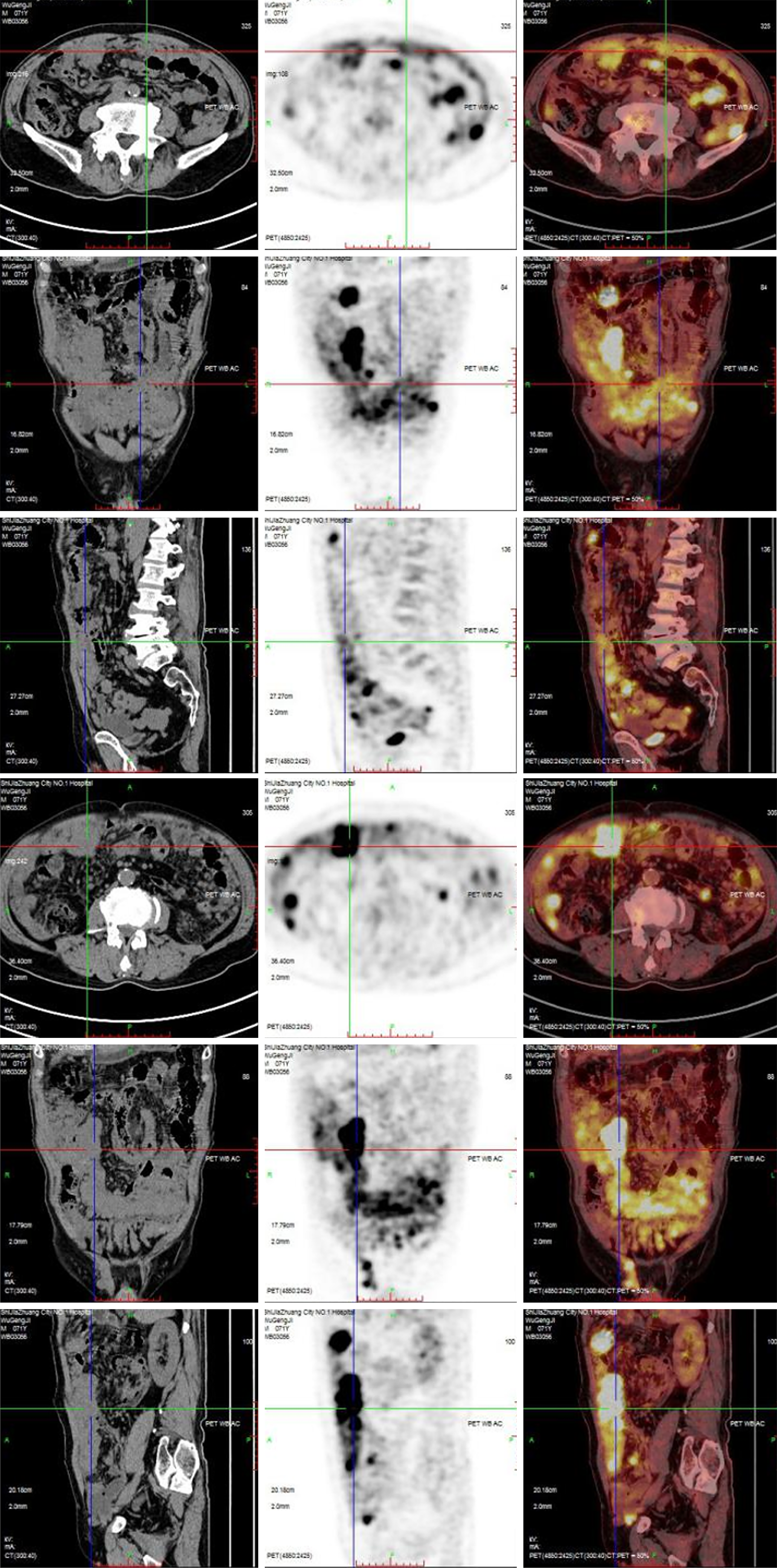
Figure 2 Positron emission tomography-computed tomography.
It shows that the peritoneum is thickened with abnormal hypermetabolism, multiple mass or nodular soft tissue density images in abdominal/pelvic cavity and peritoneum; via enhancement scanning, the lesion is enhanced in mild to moderate level; multiple hypermetabolic lymph nodes in abdominal/pelvic cavity and right external ilium; and seroperitoneum.
- Citation: Xu JD, Wang Z, Zhou Q, Meng N, Zhang SM, Liu N. Extragastrointestinal stromal tumors with diffuse membranous distribution with bleeding: A case report. World J Clin Cases 2024; 12(26): 5990-5997
- URL: https://www.wjgnet.com/2307-8960/full/v12/i26/5990.htm
- DOI: https://dx.doi.org/10.12998/wjcc.v12.i26.5990


