Copyright
©The Author(s) 2024.
World J Clin Cases. Sep 16, 2024; 12(26): 5974-5982
Published online Sep 16, 2024. doi: 10.12998/wjcc.v12.i26.5974
Published online Sep 16, 2024. doi: 10.12998/wjcc.v12.i26.5974
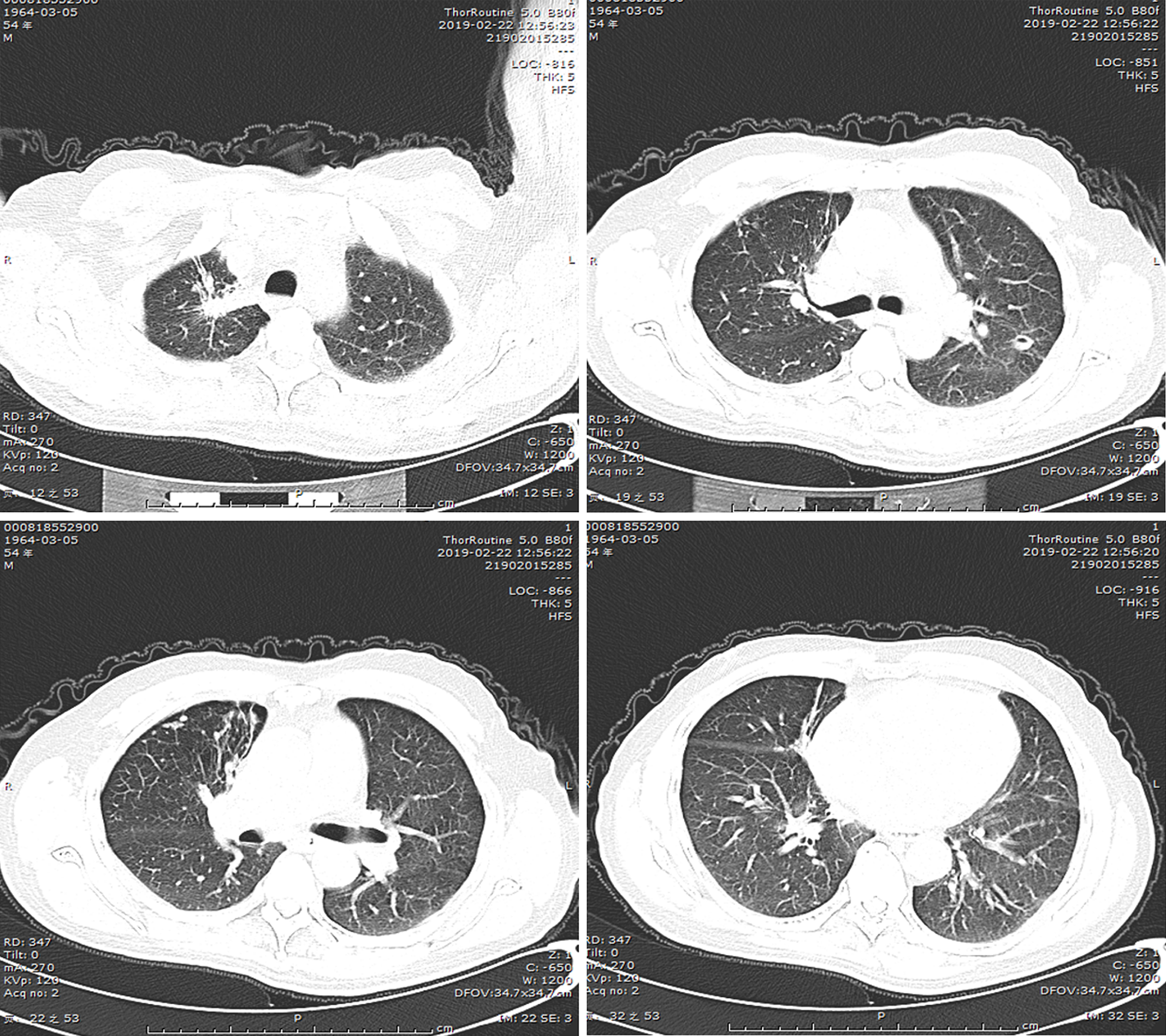
Figure 4
Following a 5-month course of anti-tuberculosis treatment, the subsequent computed tomography scan revealed complete closure of the cavity in the right upper lung, absorbtion of pleural effusion in the right hemithorax, and significant reduction in size of the cavity in the lower left lung.
- Citation: Liu M, Dong XY, Ding ZX, Wang QH, Li DH. Organizing pneumonia secondary to pulmonary tuberculosis: A case report. World J Clin Cases 2024; 12(26): 5974-5982
- URL: https://www.wjgnet.com/2307-8960/full/v12/i26/5974.htm
- DOI: https://dx.doi.org/10.12998/wjcc.v12.i26.5974


