Copyright
©The Author(s) 2024.
World J Clin Cases. Sep 16, 2024; 12(26): 5974-5982
Published online Sep 16, 2024. doi: 10.12998/wjcc.v12.i26.5974
Published online Sep 16, 2024. doi: 10.12998/wjcc.v12.i26.5974
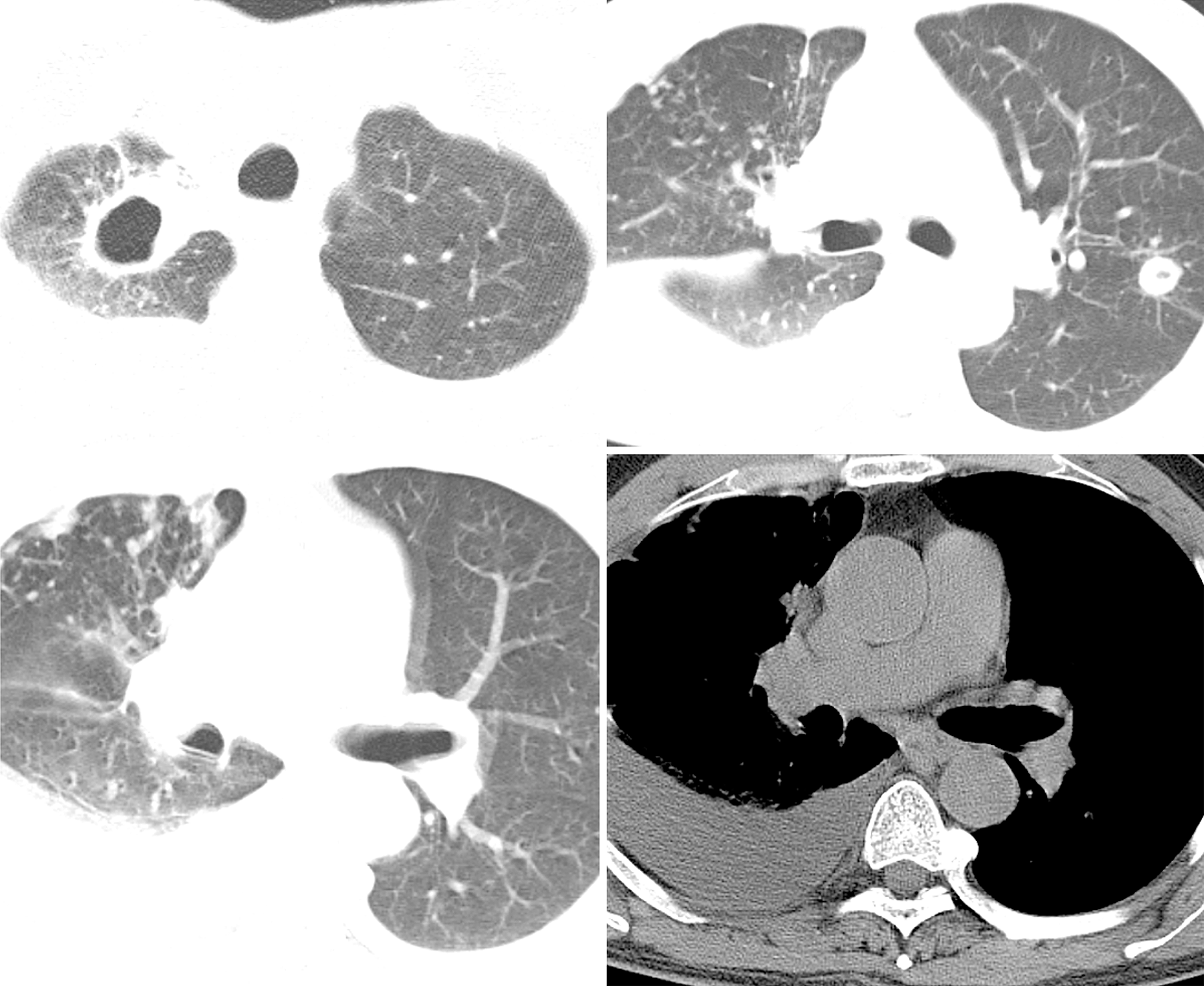
Figure 3 After admission, the computed tomography scan unveiled multiple nodules and cavities in both lungs, suggesting infection, particularly tuberculosis.
Right pleurisy and pleural effusion were noted.
- Citation: Liu M, Dong XY, Ding ZX, Wang QH, Li DH. Organizing pneumonia secondary to pulmonary tuberculosis: A case report. World J Clin Cases 2024; 12(26): 5974-5982
- URL: https://www.wjgnet.com/2307-8960/full/v12/i26/5974.htm
- DOI: https://dx.doi.org/10.12998/wjcc.v12.i26.5974


