Copyright
©The Author(s) 2024.
World J Clin Cases. Sep 16, 2024; 12(26): 5968-5973
Published online Sep 16, 2024. doi: 10.12998/wjcc.v12.i26.5968
Published online Sep 16, 2024. doi: 10.12998/wjcc.v12.i26.5968
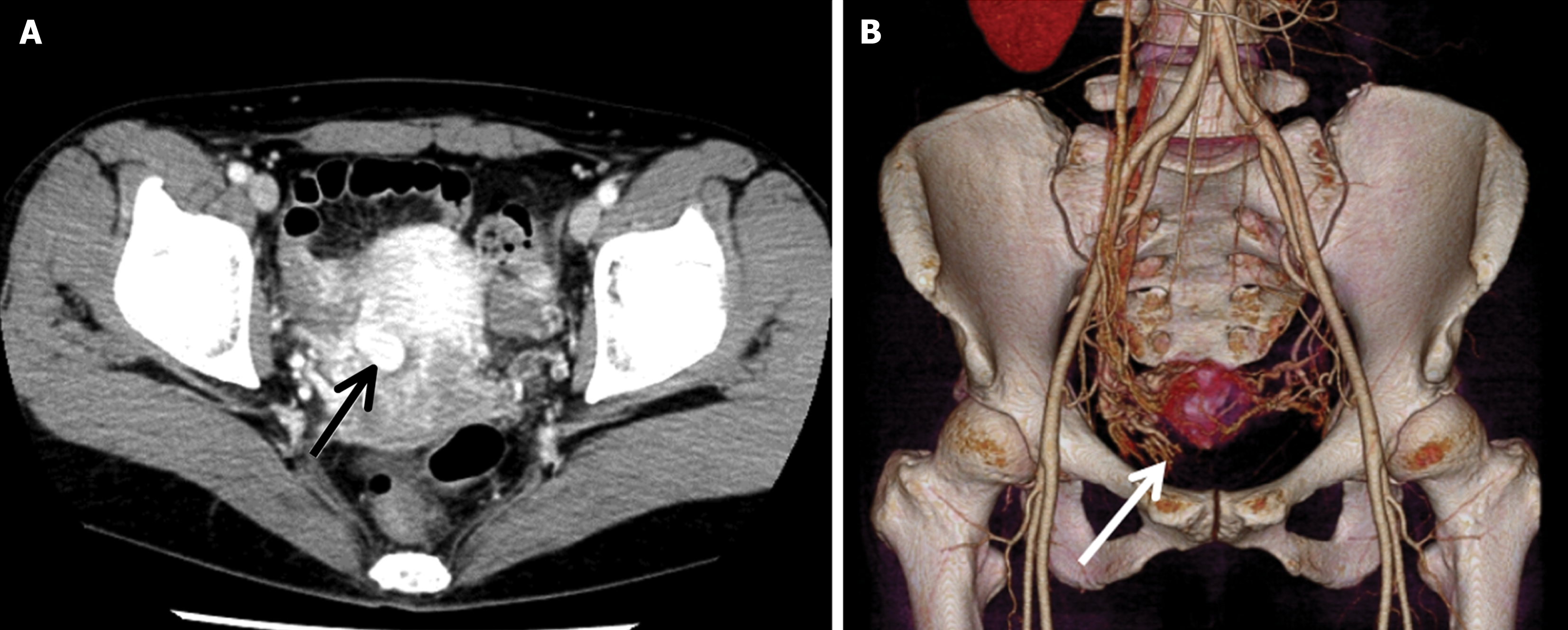
Figure 2 Pelvic contrast-enhanced computed tomography.
A: A contrast-enhanced mass (arrow) is evident in the arterial phase; a uterine artery pseudoaneurysm was diagnosed (dynamic imaging); B: Three-dimensional image reconstruction shows the uterine artery and the uterine artery pseudoaneurysm (arrow) continuous with it.
- Citation: Kakinuma K, Kakinuma T, Ueyama K, Okamoto R, Yanagida K, Takeshima N, Ohwada M. Uterine artery pseudoaneurysm caused by hysteroscopic surgery: A case report. World J Clin Cases 2024; 12(26): 5968-5973
- URL: https://www.wjgnet.com/2307-8960/full/v12/i26/5968.htm
- DOI: https://dx.doi.org/10.12998/wjcc.v12.i26.5968


