Copyright
©The Author(s) 2024.
World J Clin Cases. Sep 6, 2024; 12(25): 5775-5783
Published online Sep 6, 2024. doi: 10.12998/wjcc.v12.i25.5775
Published online Sep 6, 2024. doi: 10.12998/wjcc.v12.i25.5775
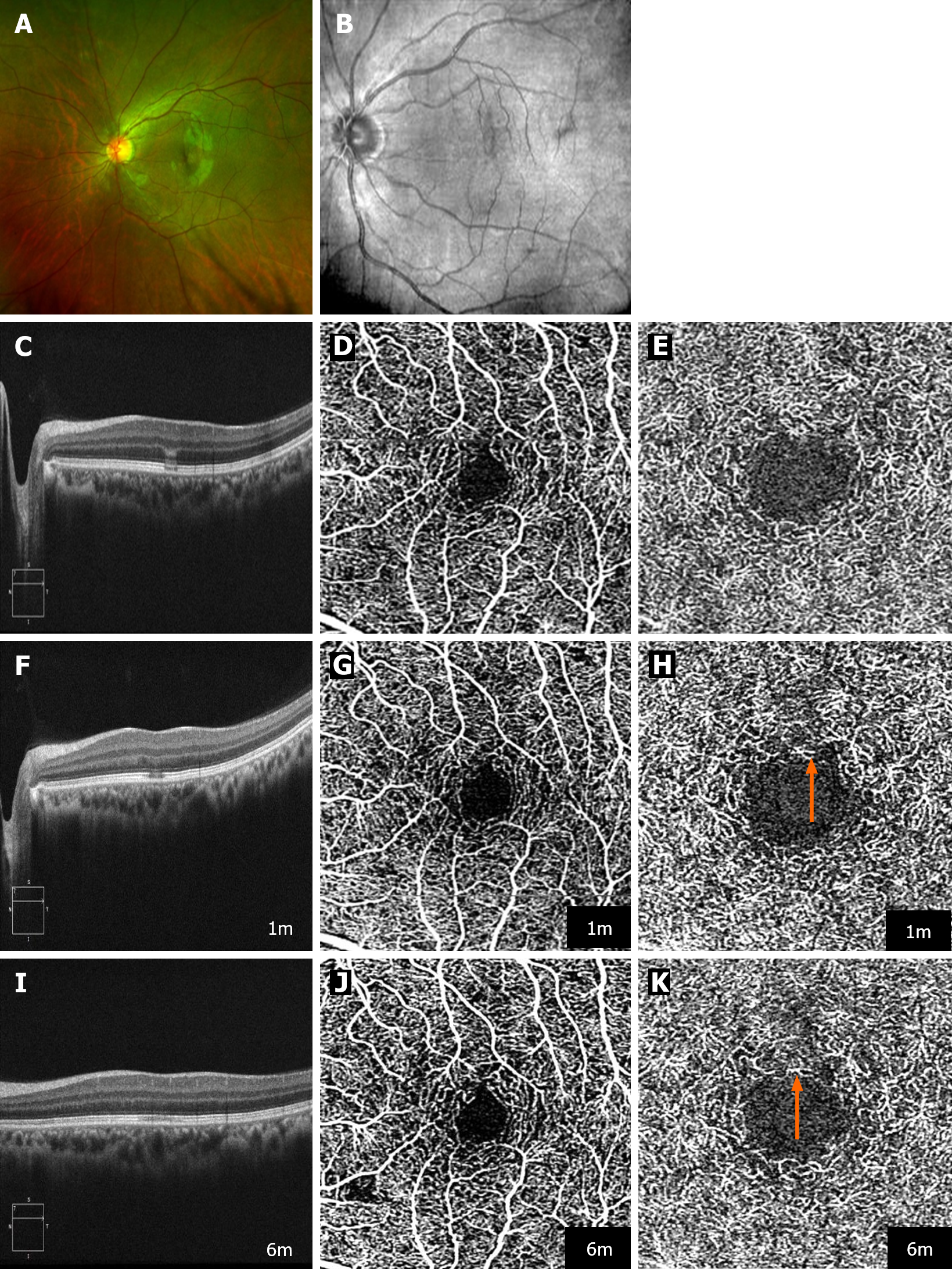
Figure 1 Multimodal imaging of the evolution of an acute macular neuroretinopathy lesion in the left eye of Case 1.
A: Ultra widefield pseudocolor fundus photography (Optos PLC, Dunfermline, Scotland, United Kingdom) shows a rugby ball-like lesion within the macular region; B: Near-infrared reflectance imaging of a hyporeflective lesion directed towards the macula; C, F and I: Sequential optical coherence tomography (OCT) B-scans shows the progression of the lesion. Initially, the images show a hyporeflective external limiting membrane, a hyperreflective myoid zone, and a hyporeflective ellipsoid zone (EZ), alongside the collapse of the outer plexiform layer with hyperreflective Henle's fiber layer and loss of the interdigitation zone (IZ). Follow-up at 1 mon reveals persistence of the hyporeflective EZ and deterioration in the IZ, which remains the sole impairment after 6 mon; D, G and J: Tracked OCT-angiography (OCTA) (Plex Elite 9000; Carl Zeiss Meditec, Inc., Dublin, CA, United States), targeting the superficial capillary plexus, indicating no observable flow signal deficits throughout the follow-up period; E, H and K: Tracked OCTA (Plex Elite 9000; Carl Zeiss Meditec, Inc.) at the deep capillary plexus level, showing an emergence and subsequent exacerbation of flow signal deficits (indicated by orange arrow) at 1 mo and 6 mo.
- Citation: Bi C, Huang CM, Shi YQ, Huang C, Yu T. Acute macular neuroretinopathy following COVID-19 infection: Three case reports. World J Clin Cases 2024; 12(25): 5775-5783
- URL: https://www.wjgnet.com/2307-8960/full/v12/i25/5775.htm
- DOI: https://dx.doi.org/10.12998/wjcc.v12.i25.5775


