Copyright
©The Author(s) 2024.
World J Clin Cases. Sep 6, 2024; 12(25): 5761-5768
Published online Sep 6, 2024. doi: 10.12998/wjcc.v12.i25.5761
Published online Sep 6, 2024. doi: 10.12998/wjcc.v12.i25.5761
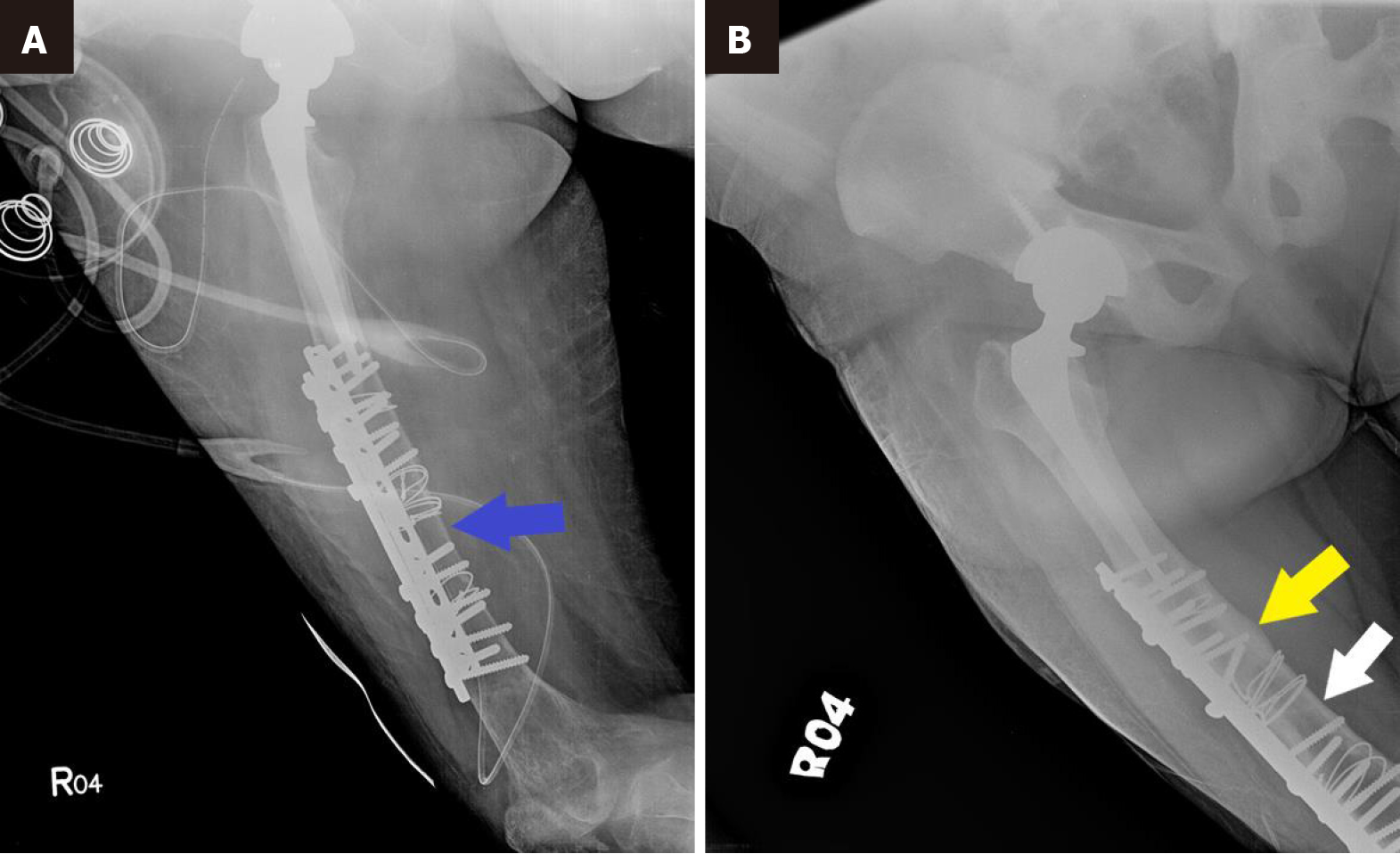
Figure 6 Hip X-rays.
A: Hip X-ray shows thin cortical bone (blue arrow) at the time the fracture occurred; B: Hip X-ray 3 years after subtotal parathyroidectomy and 2 years after right hip replacement shows solid union of the fracture (yellow arrow), with restoration of the cortical bone thickness (white arrow).
- Citation: Lin TC, Lin SW, Yeh KT. Parathyroidectomy restored bone mineral density in a neglected femoral neck fracture with renal osteodystrophy: A case report. World J Clin Cases 2024; 12(25): 5761-5768
- URL: https://www.wjgnet.com/2307-8960/full/v12/i25/5761.htm
- DOI: https://dx.doi.org/10.12998/wjcc.v12.i25.5761


