Copyright
©The Author(s) 2024.
World J Clin Cases. Aug 26, 2024; 12(24): 5596-5603
Published online Aug 26, 2024. doi: 10.12998/wjcc.v12.i24.5596
Published online Aug 26, 2024. doi: 10.12998/wjcc.v12.i24.5596
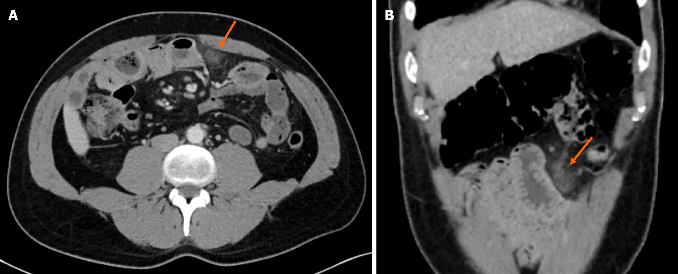
Figure 3 Omental infarction in a 34-year-old man.
A and B: On contrast enhanced axial (A) and coronal (B) computed tomography images, a mass like focal hyperdensity with peripheral fat stranding was seen in the left inframesocolic omentum (arrows).
- Citation: Kar H, Khabbazazar D, Acar N, Karasu Ş, Bağ H, Cengiz F, Dilek ON. Are all primary omental infarcts truly idiopathic? Five case reports. World J Clin Cases 2024; 12(24): 5596-5603
- URL: https://www.wjgnet.com/2307-8960/full/v12/i24/5596.htm
- DOI: https://dx.doi.org/10.12998/wjcc.v12.i24.5596


