Copyright
©The Author(s) 2024.
World J Clin Cases. Aug 26, 2024; 12(24): 5589-5595
Published online Aug 26, 2024. doi: 10.12998/wjcc.v12.i24.5589
Published online Aug 26, 2024. doi: 10.12998/wjcc.v12.i24.5589
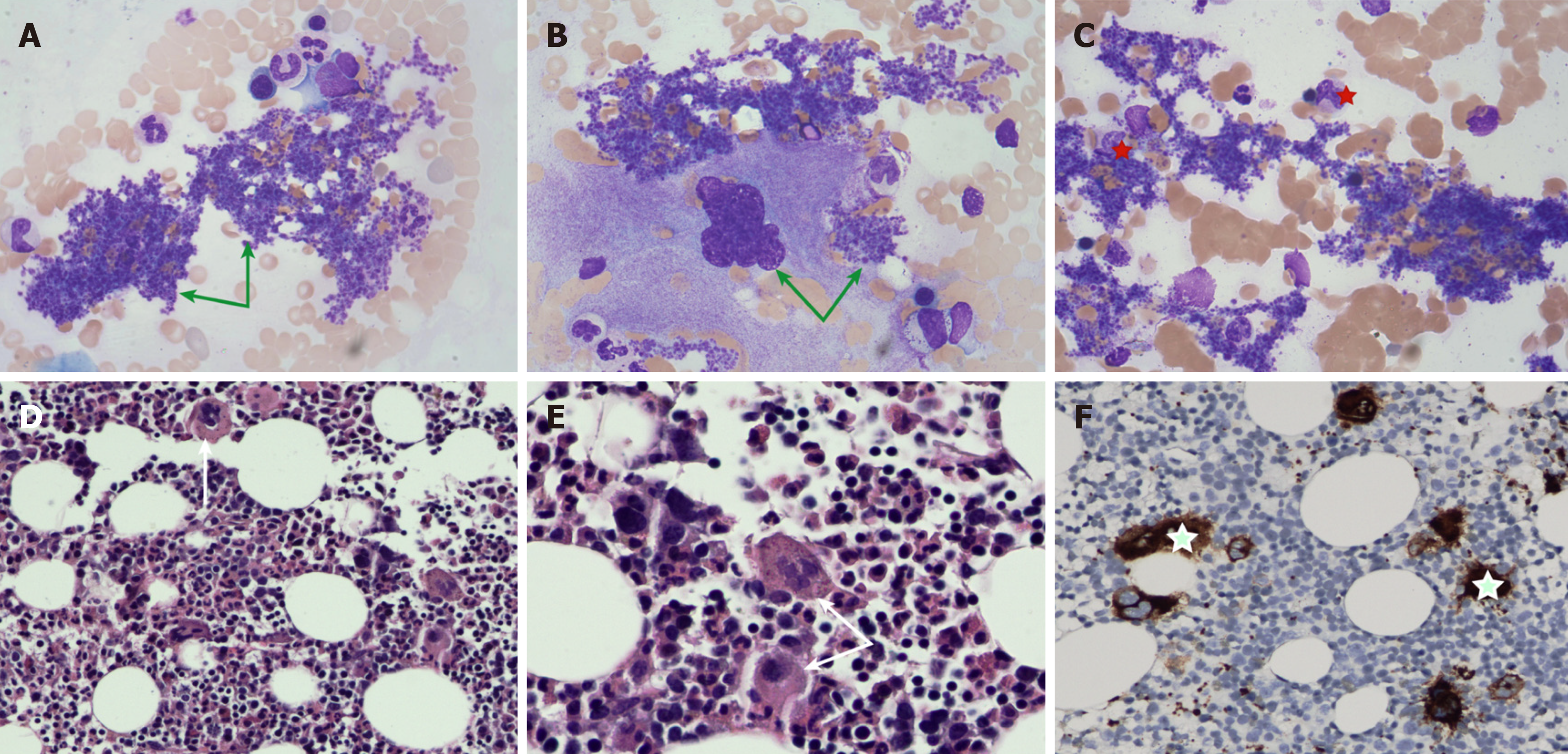
Figure 2 Bone marrow examination.
A and B: Platelets existed in the form of piles and pieces in the peripheral blood smear and bone marrow smear (green arrows); C: Proplatelet-producing megakaryocytes were markedly increased in the bone marrow smear (red five-pointed star); D and E: The number of megakaryocytes was significantly elevated, especially megakaryocytes with large cell bodies and hyperlobated nuclei, and they were isolated or arranged in dense clusters (reticular fibrosis grade of MF-1) (Hematoxylin-eosin staining: 200 times, 400 times) (white arrows); F: CD61 immunohistochemistry was also positive on megakaryocytes (green five-pointed star).
- Citation: Wu ZN, JI R, Xiao Y, Wang YD, Zhao CY. IgG4-related sclerosing cholangitis associated with essential thrombocythemia: A case report. World J Clin Cases 2024; 12(24): 5589-5595
- URL: https://www.wjgnet.com/2307-8960/full/v12/i24/5589.htm
- DOI: https://dx.doi.org/10.12998/wjcc.v12.i24.5589


