Copyright
©The Author(s) 2024.
World J Clin Cases. Aug 16, 2024; 12(23): 5431-5440
Published online Aug 16, 2024. doi: 10.12998/wjcc.v12.i23.5431
Published online Aug 16, 2024. doi: 10.12998/wjcc.v12.i23.5431
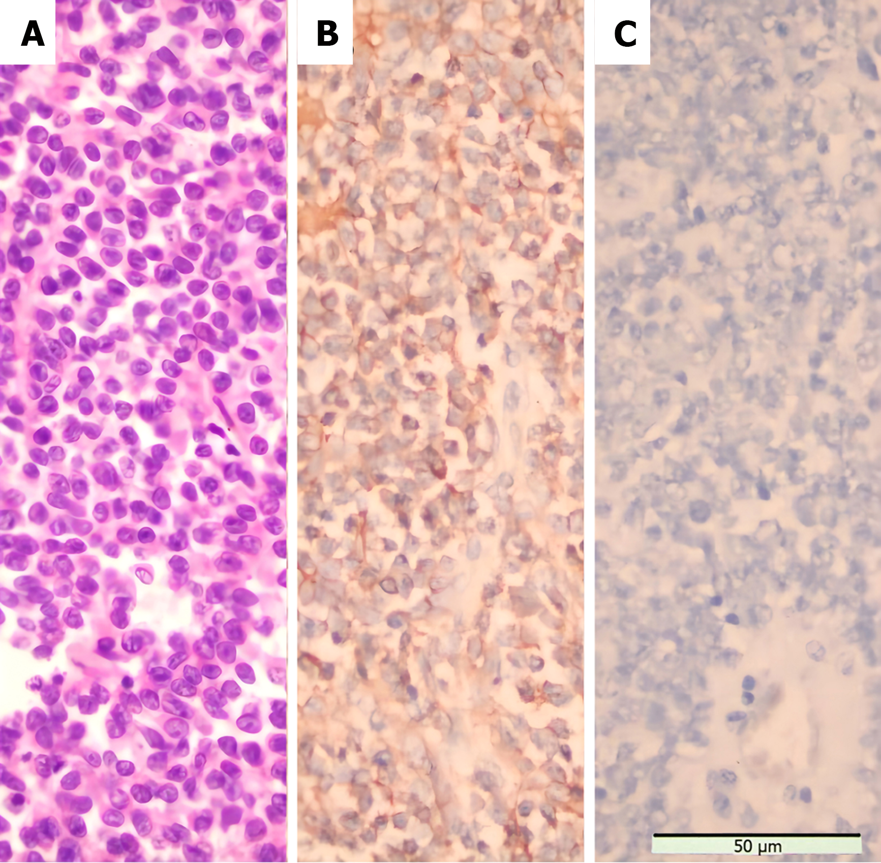
Figure 6 Morphological features and immunostaining results of the tumor.
A: Small, round tumor cells can be seen scattered throughout the visual field Staining method: HE staining (400 ×); B: Diffuse hyperstaining of tumor cell membrane for CD99 Staining method: Polymer method (400 ×); C: No positive reaction was observed for adrenocor ticotropic hormore staining method: Polymer method (400 ×).
- Citation: Dong GF, Hou YK, Ma Q, Ma SY, Wang YJ, Rexiati M, Wang WG. Cushing's syndrome caused by giant Ewing's sarcoma of the kidney: A case report and review of literature. World J Clin Cases 2024; 12(23): 5431-5440
- URL: https://www.wjgnet.com/2307-8960/full/v12/i23/5431.htm
- DOI: https://dx.doi.org/10.12998/wjcc.v12.i23.5431


