Copyright
©The Author(s) 2024.
World J Clin Cases. Aug 16, 2024; 12(23): 5431-5440
Published online Aug 16, 2024. doi: 10.12998/wjcc.v12.i23.5431
Published online Aug 16, 2024. doi: 10.12998/wjcc.v12.i23.5431
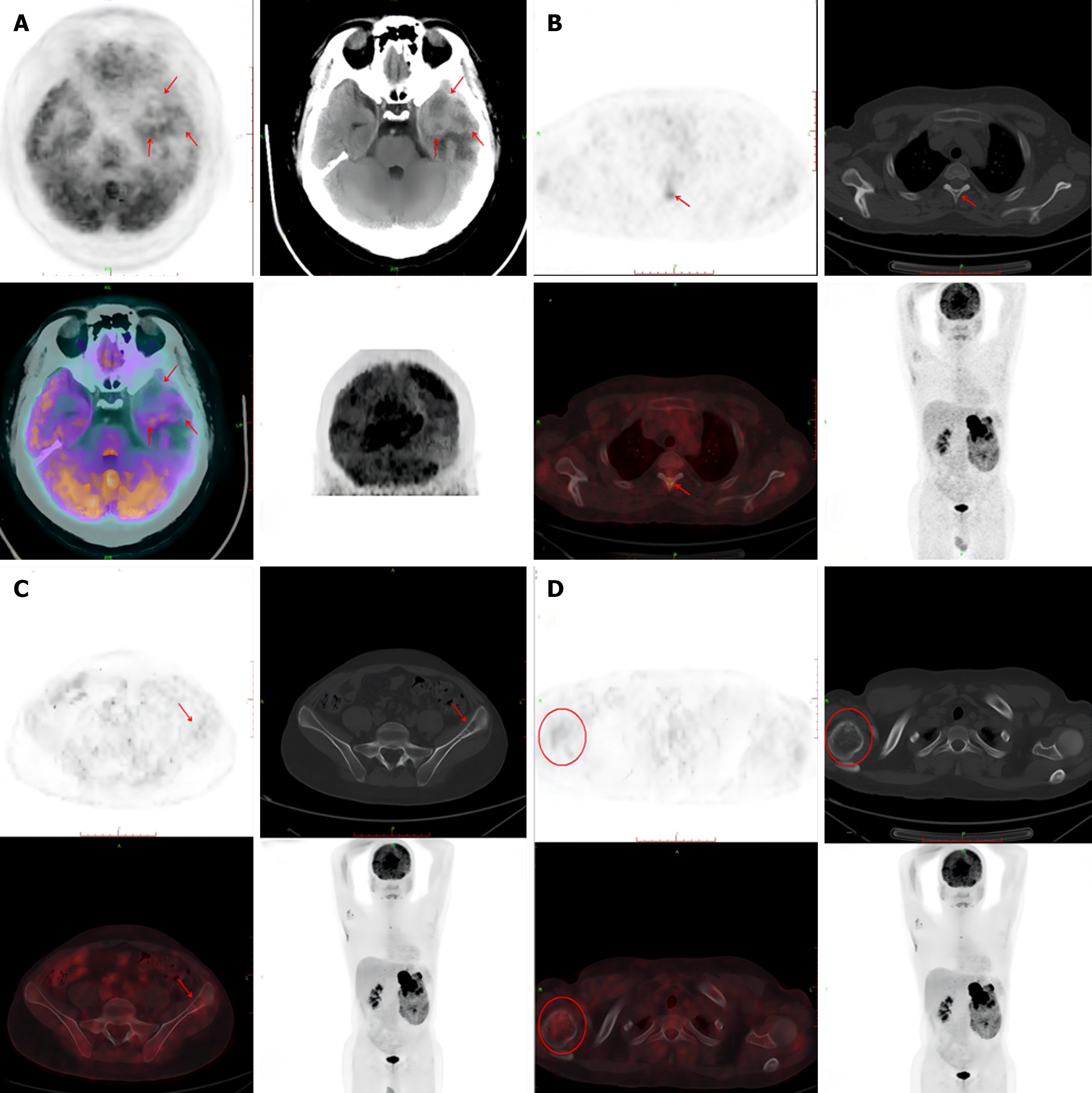
Figure 4 Nuclear medicine image of metastatic tumors.
A: The shape of the brain was as usual, with a slightly high-density mass in the left temporal pole and a few low-density shadows in the center. The larger section was about 3.3 cm × 2.2 cm. The periphery of the lesion showed large patches of low-density edema. PET showed annular high uptake of radioactivity in the lesion, Maximum Standardized Uptake Value (SUVmax) 9.1; B: High radioactive uptake shadow was found in the thoracic third vertebral arch plate, SUVmax 5.0, and no obvious bone destruction was found in the corresponding part; C and D: Left iliac wing, right humeral head and local bone of density is decreas, and a small amount of radioactive uptake was observed in the corresponding parts. The SUVmax was 2.7.
- Citation: Dong GF, Hou YK, Ma Q, Ma SY, Wang YJ, Rexiati M, Wang WG. Cushing's syndrome caused by giant Ewing's sarcoma of the kidney: A case report and review of literature. World J Clin Cases 2024; 12(23): 5431-5440
- URL: https://www.wjgnet.com/2307-8960/full/v12/i23/5431.htm
- DOI: https://dx.doi.org/10.12998/wjcc.v12.i23.5431


