Copyright
©The Author(s) 2024.
World J Clin Cases. Aug 6, 2024; 12(22): 5263-5270
Published online Aug 6, 2024. doi: 10.12998/wjcc.v12.i22.5263
Published online Aug 6, 2024. doi: 10.12998/wjcc.v12.i22.5263
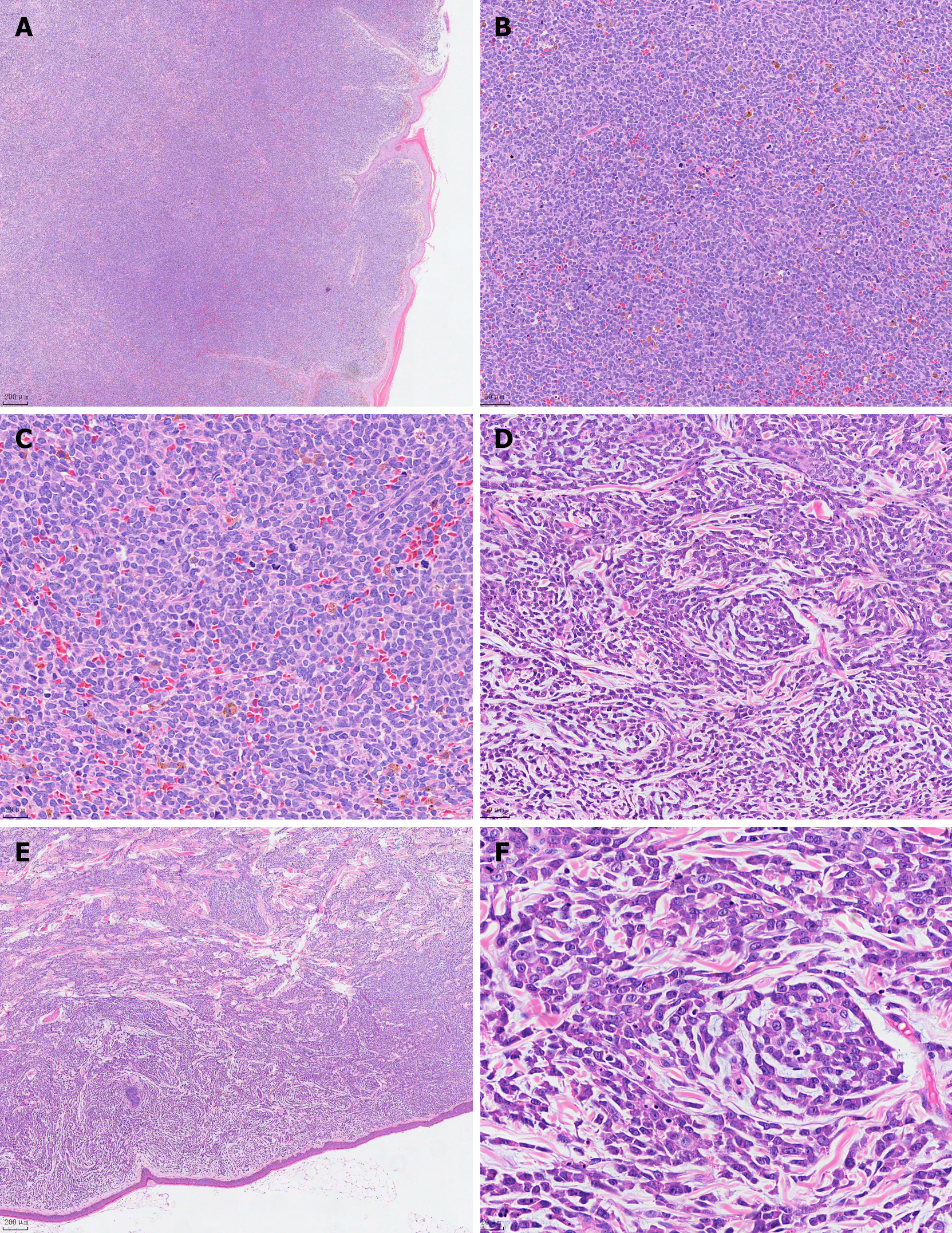
Figure 2 Microscopic pathological images of HE in two patients.
A: Case 1 tumor cells do not invade the epidermis, the cell-free zone between the epidermis (HE staining, 40 times), EnVision two-step method, Bar = 200 μm; B: Case 2 tumor cells do not invade the epidermis, interepidermal cell-free zone (HE staining, 40 times), EnVision two-step method, Bar = 200 μm; C: Case1 tumor cells were single, medium size, nuclear round or oval, chromatin fine (HE, 200 ×), EnVision two-step method, Bar = 50 μm; D: Case 2 tumor cells single, medium size, round or oval nuclei, fine chromatin (HE, 200 ×), EnVision two-step method, Bar = 50 μm; E: Case1 case image showing small nucleolus and nuclear fission (HE, 400 ×), EnVision two-step method, Bar = 20 μm; F: Case 2 case images showing small nucleolus and nuclear fission (HE, 400 ×), EnVision two-step method, Bar = 20 μm.
- Citation: Cai JW, Li MY, Wang WH, Shi HQ, Yang YH, Chen JJ. Blastic plasmacytoid dendritic cell neoplasm in Jinhua, China: Two case reports. World J Clin Cases 2024; 12(22): 5263-5270
- URL: https://www.wjgnet.com/2307-8960/full/v12/i22/5263.htm
- DOI: https://dx.doi.org/10.12998/wjcc.v12.i22.5263


