Copyright
©The Author(s) 2024.
World J Clin Cases. Aug 6, 2024; 12(22): 5258-5262
Published online Aug 6, 2024. doi: 10.12998/wjcc.v12.i22.5258
Published online Aug 6, 2024. doi: 10.12998/wjcc.v12.i22.5258
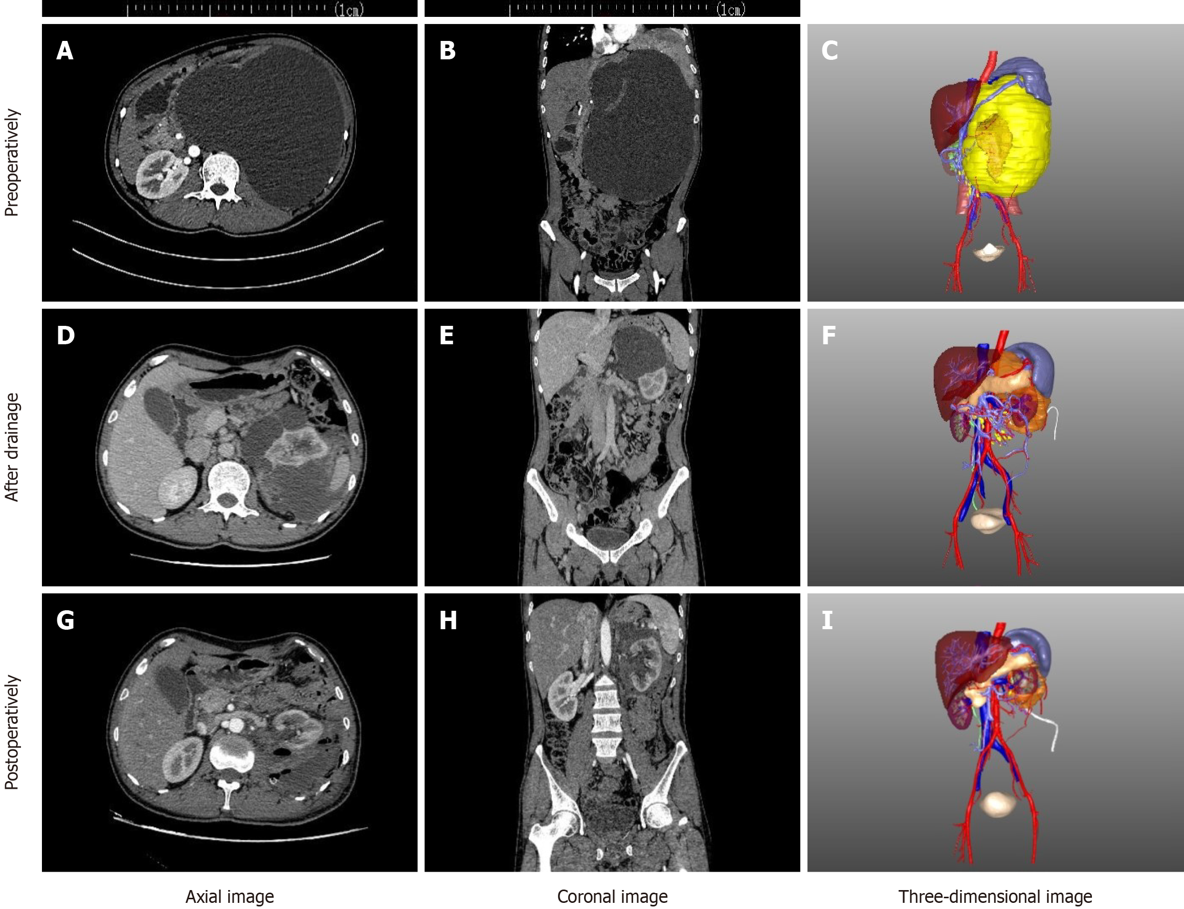
Figure 1 Computed tomography and 3D reconstruction images showing the volume reduction of the cystic mass before and after ultrasound-guided percutaneous nephrostomy.
A large cystic mass was found around the left kidney. A: Preoperative sagittal computed tomography (CT) images; B: Preoperative CT coronal images; C: Preoperative three-dimensional coronal image; D: Sagittal CT images after drainage; E: CT coronal image after drainage; F: 3D coronal image after drainage; G: Sagittal CT images after surgery; H: Postoperative CT coronal image; I: 3D coronal image after surgery.
- Citation: Wang YK, Liu YH, Shuang WB. Giant retroperitoneal hemolymphangioma: A case report and review of literature. World J Clin Cases 2024; 12(22): 5258-5262
- URL: https://www.wjgnet.com/2307-8960/full/v12/i22/5258.htm
- DOI: https://dx.doi.org/10.12998/wjcc.v12.i22.5258


