Copyright
©The Author(s) 2024.
World J Clin Cases. Aug 6, 2024; 12(22): 5217-5224
Published online Aug 6, 2024. doi: 10.12998/wjcc.v12.i22.5217
Published online Aug 6, 2024. doi: 10.12998/wjcc.v12.i22.5217
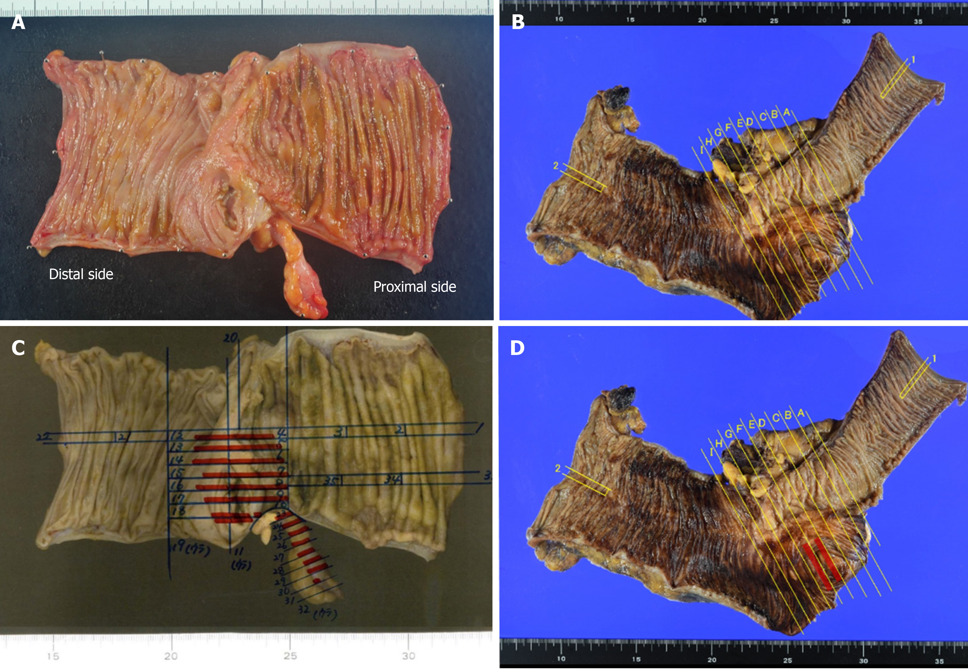
Figure 4 Resected specimen and tumor mapping.
A-C: Although no neoplastic changes are seen at the cecal surface or appendiceal orifice in Case 1 (A) or Case 4 (C), tumor cells have invaded widely into the cecum in Case 1 (B); D: Residual tumor cells are seen microscopically at the cecum around the appendiceal orifice in Case 4. Red lines show the existence of tumor cells.
- Citation: Toshima T, Inada R, Sakamoto S, Takeda E, Yoshioka T, Kumon K, Mimura N, Takata N, Tabuchi M, Oishi K, Sato T, Sui K, Okabayashi T, Ozaki K, Nakamura T, Shibuya Y, Matsumoto M, Iwata J. Goblet cell carcinoid of the appendix: Six case reports. World J Clin Cases 2024; 12(22): 5217-5224
- URL: https://www.wjgnet.com/2307-8960/full/v12/i22/5217.htm
- DOI: https://dx.doi.org/10.12998/wjcc.v12.i22.5217


