Copyright
©The Author(s) 2024.
World J Clin Cases. Aug 6, 2024; 12(22): 5196-5207
Published online Aug 6, 2024. doi: 10.12998/wjcc.v12.i22.5196
Published online Aug 6, 2024. doi: 10.12998/wjcc.v12.i22.5196
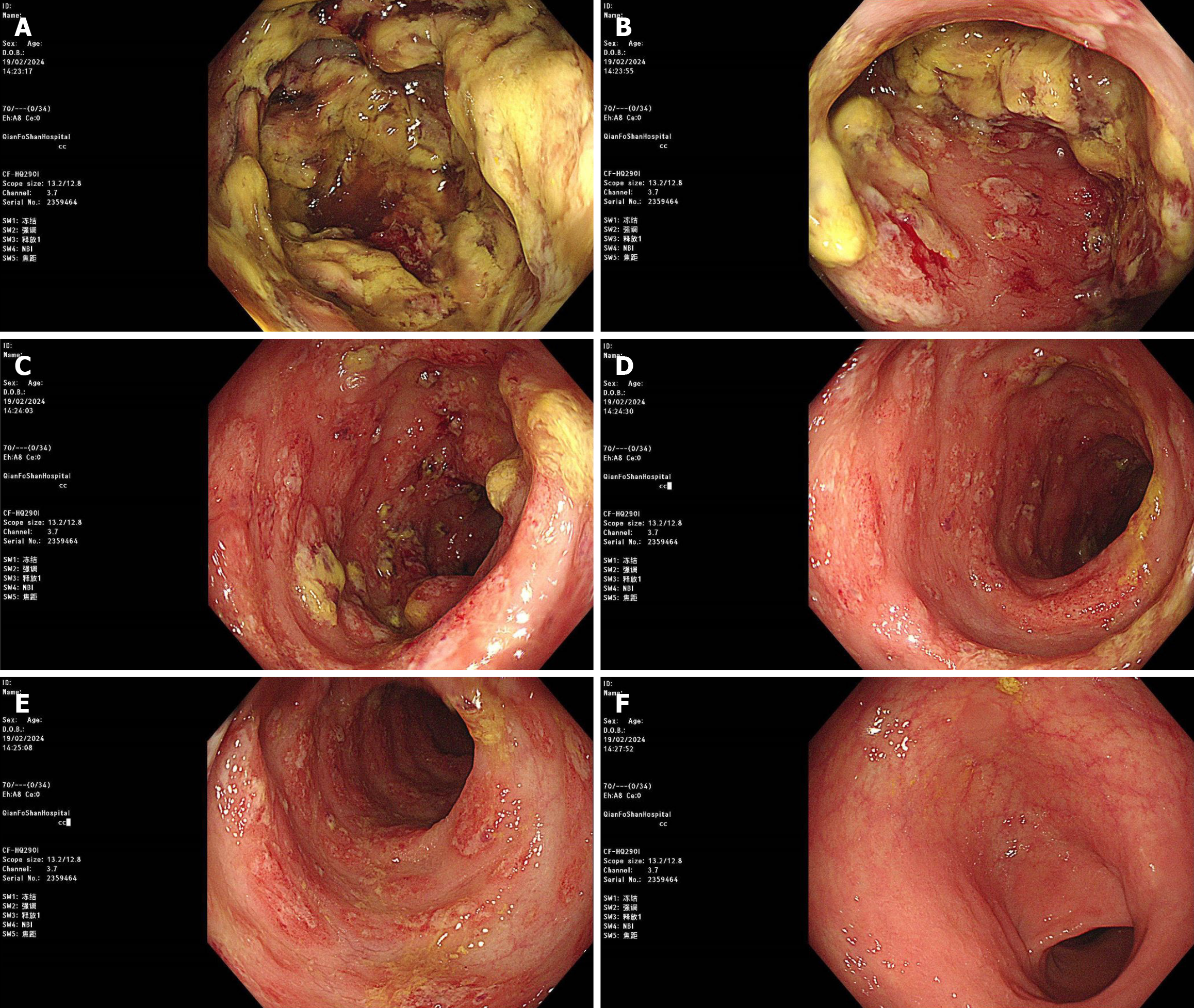
Figure 2 Colonoscopy images.
A and B: Local ulcer formation with intestinal lumen stenosis at 60 cm (A) and 55 cm (B) from the anus; C: Local mucosal edema with ulcer formation in the descending colon; D-F: Local mucosal congestion and edema with erosion in the sigmoid colon (D and E) and rectum (F).
- Citation: Yan MX. Pleural effusion, ascites, colon ulcers and hematochezia: What we can learn from the diagnostic process of a patient with plasma cell myeloma: A case report. World J Clin Cases 2024; 12(22): 5196-5207
- URL: https://www.wjgnet.com/2307-8960/full/v12/i22/5196.htm
- DOI: https://dx.doi.org/10.12998/wjcc.v12.i22.5196


