Copyright
©The Author(s) 2024.
World J Clin Cases. Jul 26, 2024; 12(21): 4783-4788
Published online Jul 26, 2024. doi: 10.12998/wjcc.v12.i21.4783
Published online Jul 26, 2024. doi: 10.12998/wjcc.v12.i21.4783
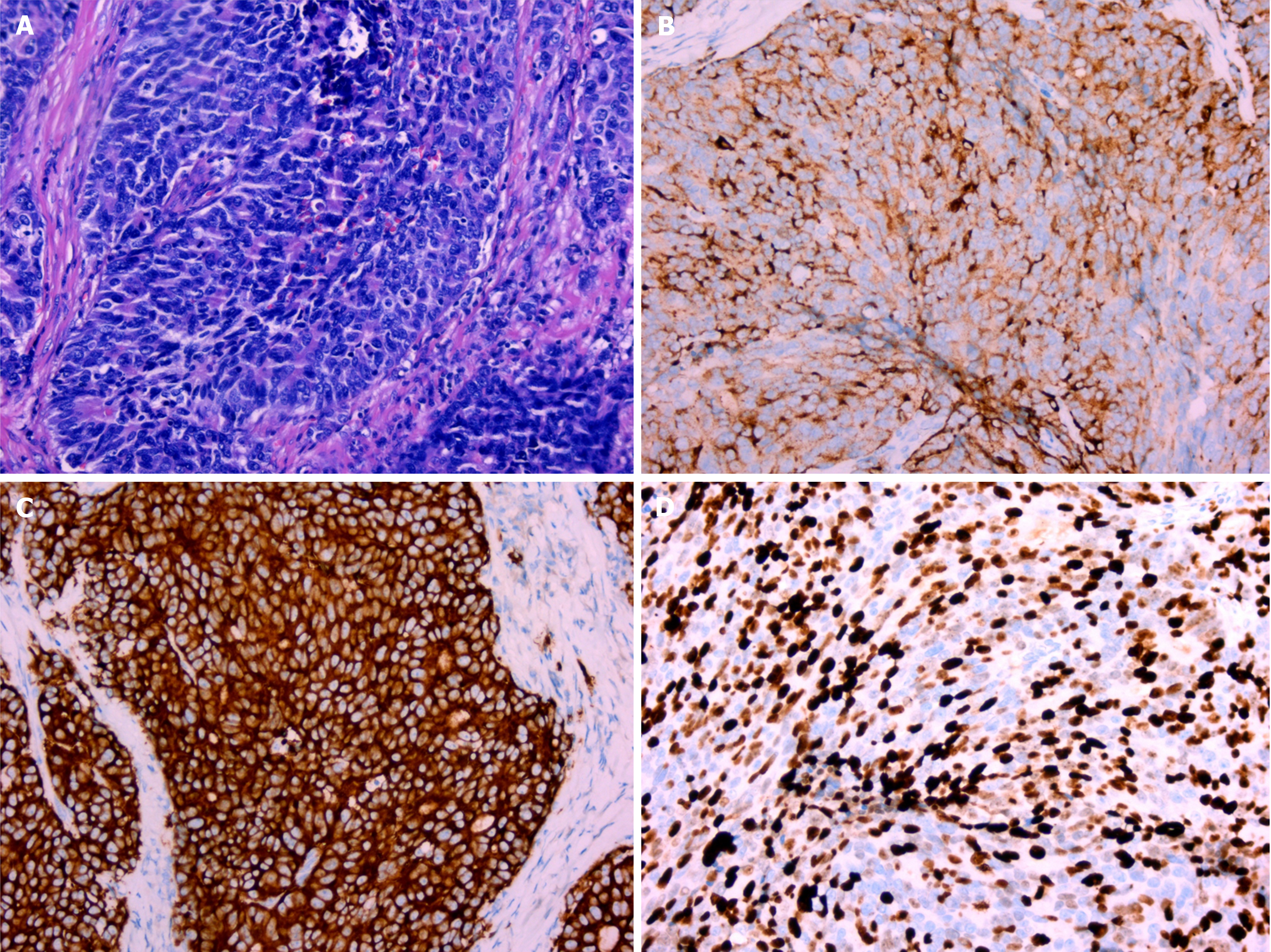
Figure 3 Pathological examination and immunohistochemical staining after surgery.
A: Hematoxylin and eosin staining revealed that the tumor cells had large nuclei, coarse granular chromatin, visible nucleoli, mitotic figures, and palisade-like structures (× 200); B: Chromogranin A was positive (× 200); C: Synaptophysin was positive (× 200); D: The Ki67 index was 70% (× 200).
- Citation: Bai LL, Guo YX, Song SY, Li R, Jiang YQ. Primary large cell neuroendocrine carcinoma of the bladder: A case report. World J Clin Cases 2024; 12(21): 4783-4788
- URL: https://www.wjgnet.com/2307-8960/full/v12/i21/4783.htm
- DOI: https://dx.doi.org/10.12998/wjcc.v12.i21.4783


