Copyright
©The Author(s) 2024.
World J Clin Cases. Jul 26, 2024; 12(21): 4770-4776
Published online Jul 26, 2024. doi: 10.12998/wjcc.v12.i21.4770
Published online Jul 26, 2024. doi: 10.12998/wjcc.v12.i21.4770
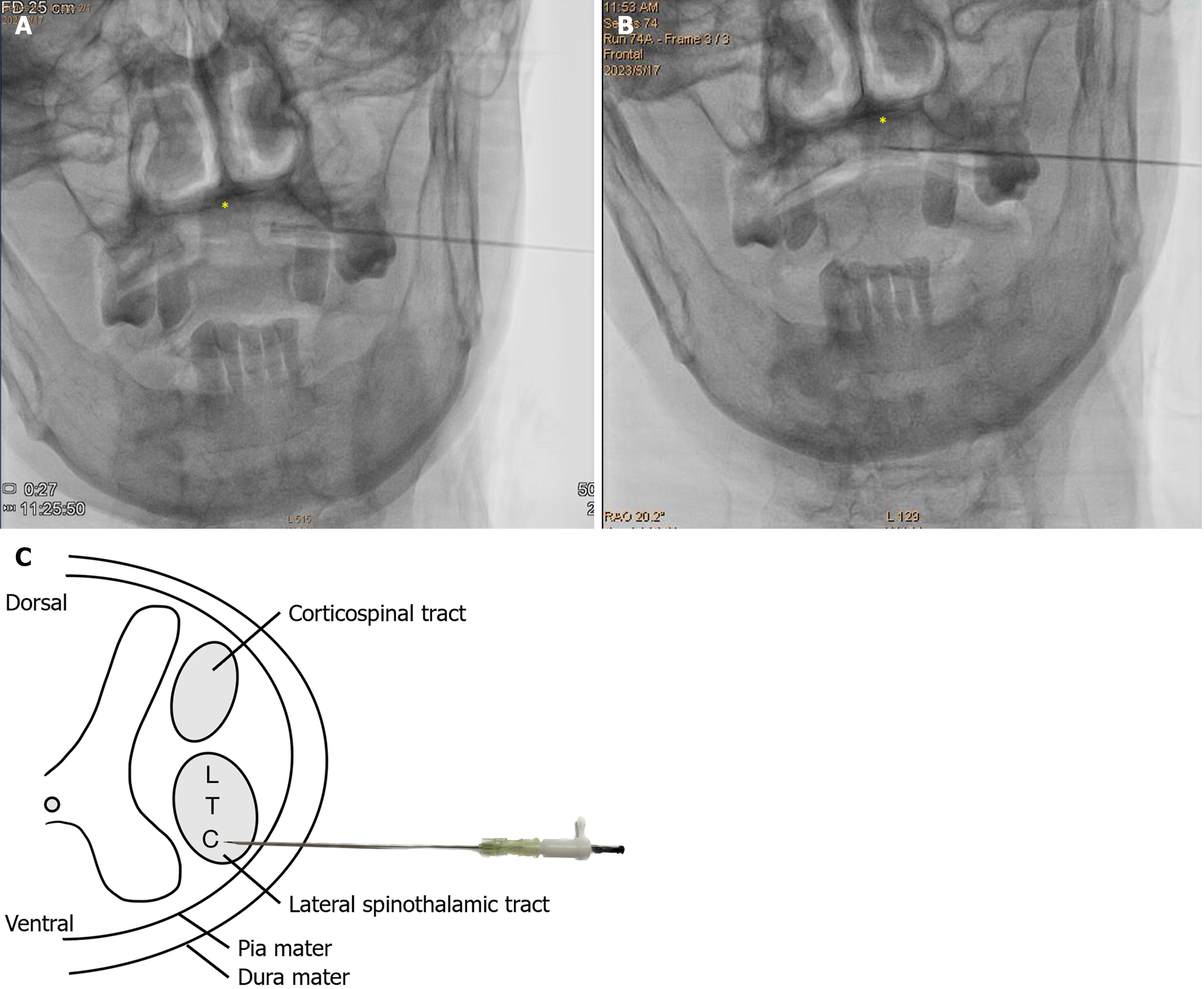
Figure 3 Fluoroscopy image of the anterior-posterior view when advancing the spinal needle during percutaneous cervical cordotomy.
A: When the needle tip enters the intrathecal space, the cerebrospinal fluid is drained. The needle tip did not cross through the midline, which is indicated by an asterisk, the dens of axis (C2); B: Radiofrequency (RF) generator was inserted into the spinal needle. The patient’s discomfort area was triggered by the sensory test. After confirmation of the target lesion, RF thermocoagulation was applied to destroy the neuropathway of the pain; C: Schematic illustration of percutaneous cervical cordotomy and the relative anatomical position of the lateral spinothalamic tract. C: Cervical; L: Lumbar; T: Thoracic.
- Citation: Lu KY, Lin FS, Lin CS, Lao HC. Percutaneous cervical cordotomy for managing refractory pain in a patient with a Pancoast tumor: A case report. World J Clin Cases 2024; 12(21): 4770-4776
- URL: https://www.wjgnet.com/2307-8960/full/v12/i21/4770.htm
- DOI: https://dx.doi.org/10.12998/wjcc.v12.i21.4770


