Copyright
©The Author(s) 2024.
World J Clin Cases. Jul 26, 2024; 12(21): 4770-4776
Published online Jul 26, 2024. doi: 10.12998/wjcc.v12.i21.4770
Published online Jul 26, 2024. doi: 10.12998/wjcc.v12.i21.4770
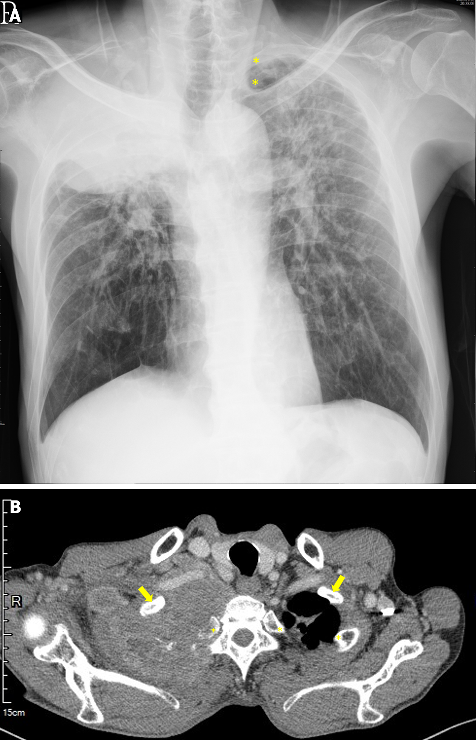
Figure 1 Preoperative imaging shows a huge space-occupying lesion at the right apical lung with adjunct bony destruction.
A: Chest radiograph. The asterisks indicate the second and third ribs at the left side, which was blurred at the right side; B: Chest computed tomography. A soft tissue mass (> 10 cm in diameter) occupied the entire right apical lung. The arrows indicate the first rib. The first rib in the right side was surrounded by the mass. The asterisk indicates the second rib. The second rib in the right side was damaged. The feature of the image highly suggested a locally advanced bronchogenic carcinoma.
- Citation: Lu KY, Lin FS, Lin CS, Lao HC. Percutaneous cervical cordotomy for managing refractory pain in a patient with a Pancoast tumor: A case report. World J Clin Cases 2024; 12(21): 4770-4776
- URL: https://www.wjgnet.com/2307-8960/full/v12/i21/4770.htm
- DOI: https://dx.doi.org/10.12998/wjcc.v12.i21.4770


