Copyright
©The Author(s) 2024.
World J Clin Cases. Jul 16, 2024; 12(20): 4412-4418
Published online Jul 16, 2024. doi: 10.12998/wjcc.v12.i20.4412
Published online Jul 16, 2024. doi: 10.12998/wjcc.v12.i20.4412
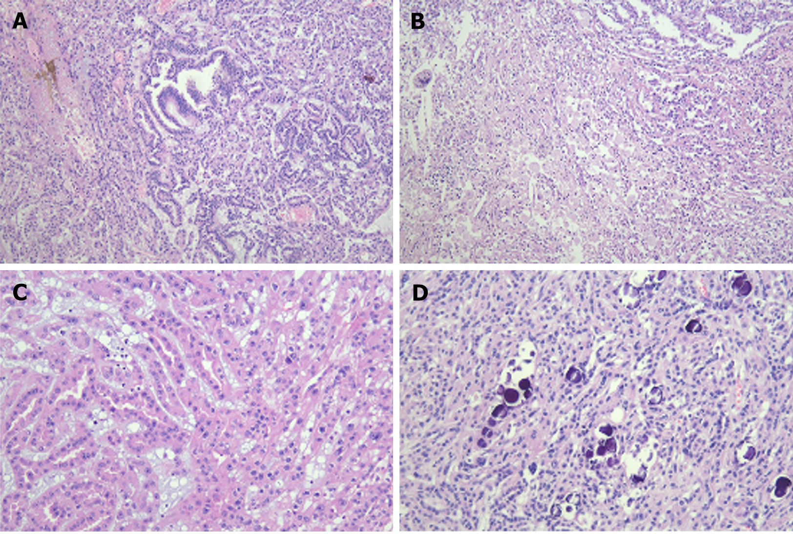
Figure 4 Microscopic findings of hematoxylin and eosin staining.
A: Tubulopapillary structures with some microcystic changes (×100); B: Papillary structures associated with accumulation of foamy histiocytes (×100); C: Tubulopapillary structures lined by eosinophilic epithelial cells with high-grade nuclear features of Fuhrman nuclear grade 3-4 (×200); D: Psammomatous calcific bodies scattered over tubular lumina and interstitial stroma (×200).
- Citation: Kim TN, Kim A, Kim KB, Lee CH. Ipsilateral retroperitoneal papillary renal cell carcinoma 27 years after simple nephrectomy for a renal abscess: A case report. World J Clin Cases 2024; 12(20): 4412-4418
- URL: https://www.wjgnet.com/2307-8960/full/v12/i20/4412.htm
- DOI: https://dx.doi.org/10.12998/wjcc.v12.i20.4412


