Copyright
©The Author(s) 2024.
World J Clin Cases. Jul 16, 2024; 12(20): 4412-4418
Published online Jul 16, 2024. doi: 10.12998/wjcc.v12.i20.4412
Published online Jul 16, 2024. doi: 10.12998/wjcc.v12.i20.4412
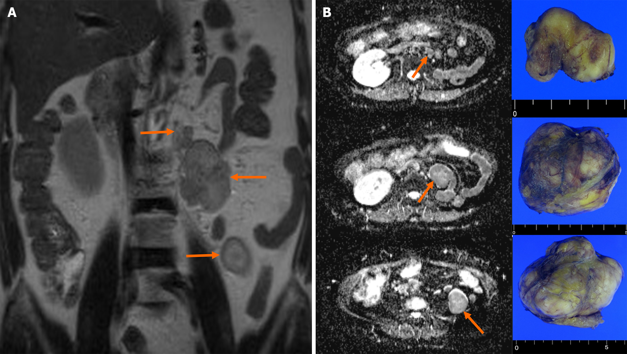
Figure 1 Magnetic resonance imaging of the abdomen.
A: The T2-weighted coronal image shows three distinct oval masses in the left retroperitoneum (indicated by orange arrows), exhibiting a high signal intensity and a well-enhanced pattern; B: The image on the left displays diffusion-weighted transverse images of three retroperitoneal masses (indicated by orange arrows), while the image on the right shows the corresponding gross features of three specimens. The diffusion-weighted transverse images reveal mild restricted diffusion.
- Citation: Kim TN, Kim A, Kim KB, Lee CH. Ipsilateral retroperitoneal papillary renal cell carcinoma 27 years after simple nephrectomy for a renal abscess: A case report. World J Clin Cases 2024; 12(20): 4412-4418
- URL: https://www.wjgnet.com/2307-8960/full/v12/i20/4412.htm
- DOI: https://dx.doi.org/10.12998/wjcc.v12.i20.4412


