Copyright
©The Author(s) 2024.
World J Clin Cases. Jul 16, 2024; 12(20): 4357-4364
Published online Jul 16, 2024. doi: 10.12998/wjcc.v12.i20.4357
Published online Jul 16, 2024. doi: 10.12998/wjcc.v12.i20.4357
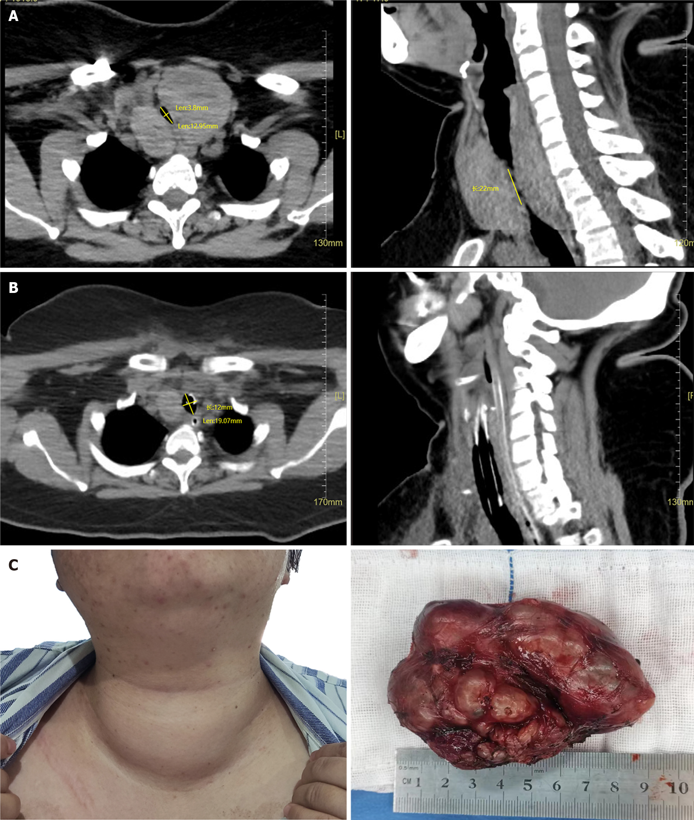
Figure 1 Pre- and postoperative neck computed tomography images and thyroid mass morphology.
A: Preoperative computed tomography (CT) images of the patient's neck, cross-sections on the left and the sagittal plane on the right; B: Post-surgery CT images of the patient's neck, cross-sections on the left and the sagittal plane on the right; C: The patient's thyroid gland before surgery (left) and post-subtotal thyroidectomy specimen of left lobe (right), right lobe specimens can be seen in supplemental figures.
- Citation: Chen XM, Jiang ZL, Wu X, Li XG. Lithium carbonate-induced giant goiter and subclinical hyperthyroidism in a patient with schizophrenia: A case report and review of literature. World J Clin Cases 2024; 12(20): 4357-4364
- URL: https://www.wjgnet.com/2307-8960/full/v12/i20/4357.htm
- DOI: https://dx.doi.org/10.12998/wjcc.v12.i20.4357


