Copyright
©The Author(s) 2024.
World J Clin Cases. Jul 16, 2024; 12(20): 4337-4347
Published online Jul 16, 2024. doi: 10.12998/wjcc.v12.i20.4337
Published online Jul 16, 2024. doi: 10.12998/wjcc.v12.i20.4337
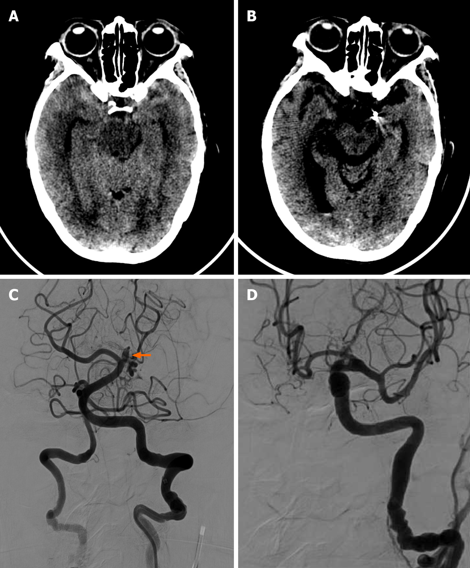
Figure 2 Case 2.
A: Axial brain computed tomography of a 79-year-old female showed bilateral subarachnoid hemorrhage in the bilateral temporal region; B: Axial brain computed tomography post-treatment showed the previous subarachnoid hemorrhage in the bilateral temporal region had subsided. No intracranial hemorrhage was found; C: Angiography showed a bulging aneurysm in the basilar perforated branches; D: Final angiography showed complete embolization of the aneurysm.
- Citation: Man IC, Pan TM, U KC. An unusual etiology of subarachnoid hemorrhage, basilar artery perforator aneurysms, in Macao: Three case reports and review of literature. World J Clin Cases 2024; 12(20): 4337-4347
- URL: https://www.wjgnet.com/2307-8960/full/v12/i20/4337.htm
- DOI: https://dx.doi.org/10.12998/wjcc.v12.i20.4337


