Copyright
©The Author(s) 2024.
World J Clin Cases. Jul 16, 2024; 12(20): 4230-4238
Published online Jul 16, 2024. doi: 10.12998/wjcc.v12.i20.4230
Published online Jul 16, 2024. doi: 10.12998/wjcc.v12.i20.4230
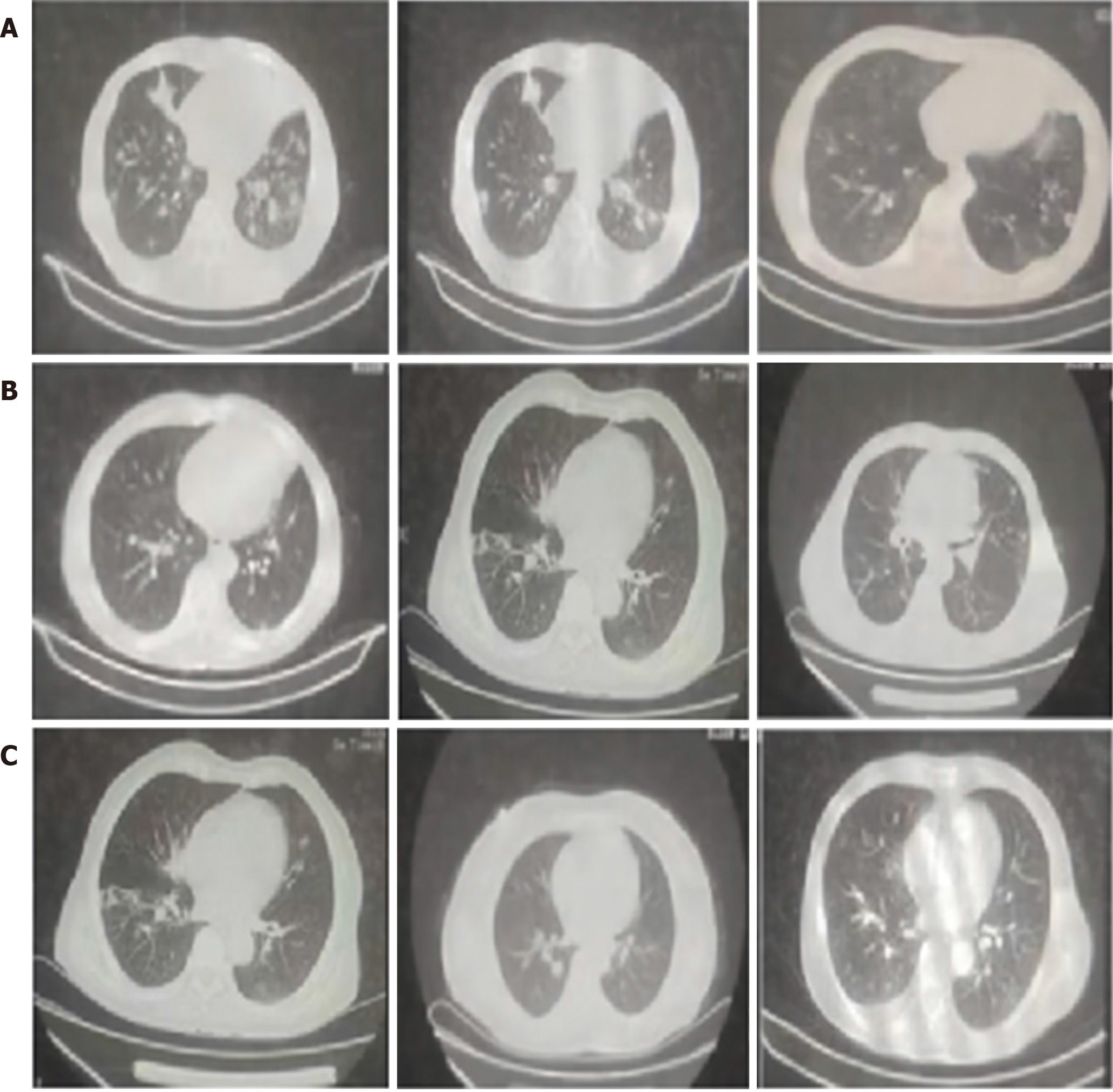
Figure 1 Pulmonary computed tomography findings of pulmonary tuberculosis and diabetes are common.
A: The patient's lungs show widespread involvement with multiple cavities of irregular shapes, with the largest cavity in the lower lung segment, with a diameter of 1.3 cm × 1.4 cm; B: Both lungs show cavities, with the left lung displaying a significant intracavitary attachment, forming a distinct crescent-shaped lucency indicative of aspergilloma formation. The right lung cavity is surrounded by slightly blurred lesions due to exudative changes, as shown in the second row; C: The influence of blood sugar levels can lead to specific pulmonary radiographic findings, such as dissemination along the bronchi. Concentrated on the right upper segment bronchial cord walking.
- Citation: Rong XS, Yao C. Computed tomography imaging and clinical significance of bacterium-positive pulmonary tuberculosis complicated with diabetes. World J Clin Cases 2024; 12(20): 4230-4238
- URL: https://www.wjgnet.com/2307-8960/full/v12/i20/4230.htm
- DOI: https://dx.doi.org/10.12998/wjcc.v12.i20.4230


