Copyright
©The Author(s) 2024.
World J Clin Cases. Jul 16, 2024; 12(20): 4154-4165
Published online Jul 16, 2024. doi: 10.12998/wjcc.v12.i20.4154
Published online Jul 16, 2024. doi: 10.12998/wjcc.v12.i20.4154
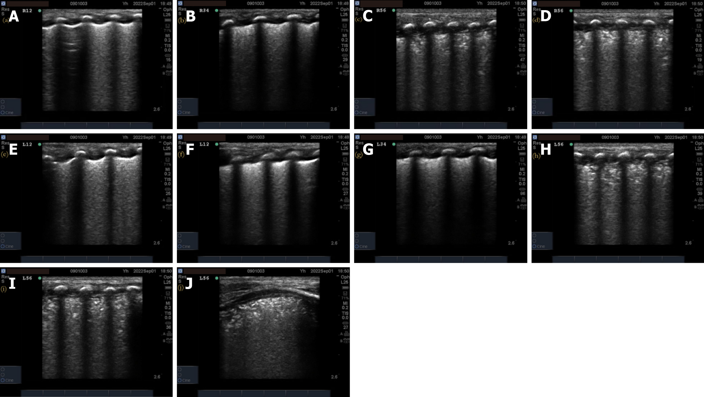
Figure 4 X-ray grade 3 respiratory distress syndrome neonatal lung ultrasonography images.
A: Ground-glass-like changes in the right lung R1–R2, as shown by the A-line, resulting in 2 points; B, E–G: Representative images showing consolidation of the left lung L1–L4 and right lung L3–L4 in half of the lung field, with 5 points for each area; C, D, H–J: Representative images showing consolidation of R5–R6/L5–L6 involving the entire lung field, with 6 points for each area and a total ultrasonography score of 29 points.
- Citation: Yang H, Gao LJ, Lei J, Li Q, Cui L, Li XH, Yin WX, Tian SH. Relationship between neonatal respiratory distress syndrome pulmonary ultrasonography and respiratory distress score, oxygenation index, and chest radiography grading. World J Clin Cases 2024; 12(20): 4154-4165
- URL: https://www.wjgnet.com/2307-8960/full/v12/i20/4154.htm
- DOI: https://dx.doi.org/10.12998/wjcc.v12.i20.4154


