Copyright
©The Author(s) 2024.
World J Clin Cases. Jul 16, 2024; 12(20): 4154-4165
Published online Jul 16, 2024. doi: 10.12998/wjcc.v12.i20.4154
Published online Jul 16, 2024. doi: 10.12998/wjcc.v12.i20.4154
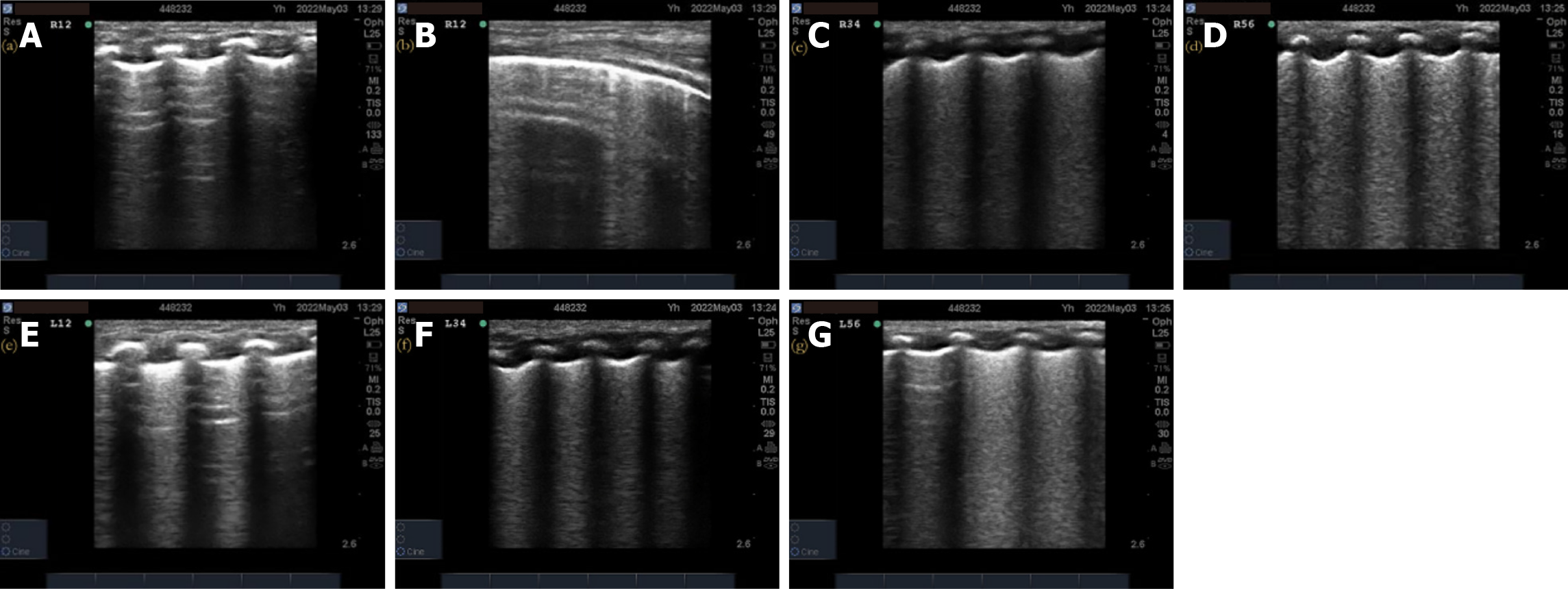
Figure 2 X-ray grade 1 respiratory distress syndrome neonatal pulmonary ultrasonography images.
A, B, E: Representative images showing increase in B-lines in the anterior chest of R1–R2/L1–L2. Real-time ultrasonography shows that B-lines and A-lines alternate with lung slip, resulting in 1 point each; C, D, F, G: Representative images showing ground-glass-like changes in each region of R3–R4/L3–L4 R5–R6/L5–L6, with 2 points each and a total ultrasonography score of 10 points.
- Citation: Yang H, Gao LJ, Lei J, Li Q, Cui L, Li XH, Yin WX, Tian SH. Relationship between neonatal respiratory distress syndrome pulmonary ultrasonography and respiratory distress score, oxygenation index, and chest radiography grading. World J Clin Cases 2024; 12(20): 4154-4165
- URL: https://www.wjgnet.com/2307-8960/full/v12/i20/4154.htm
- DOI: https://dx.doi.org/10.12998/wjcc.v12.i20.4154


