Copyright
©The Author(s) 2024.
World J Clin Cases. Jan 16, 2024; 12(2): 425-430
Published online Jan 16, 2024. doi: 10.12998/wjcc.v12.i2.425
Published online Jan 16, 2024. doi: 10.12998/wjcc.v12.i2.425
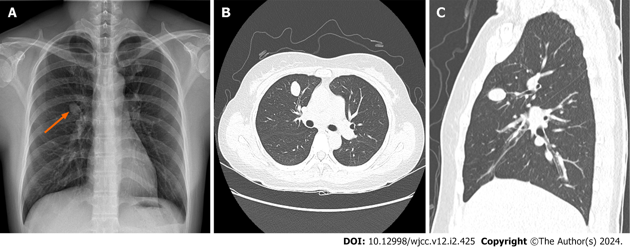
Figure 1 Preoperative imaging studies.
A: Chest radiograph showing a solitary pulmonary nodule of the right hilar area (orange arrow); B and C: Similar findings are observed on chest computed tomography (axial and sagittal views), with the nodule situated in the anterior segment of the right upper lobe.
- Citation: Ahn S, Moon Y. Uniportal video-assisted thoracoscopic fissureless right upper lobe anterior segmentectomy for inflammatory myofibroblastic tumor: A case report. World J Clin Cases 2024; 12(2): 425-430
- URL: https://www.wjgnet.com/2307-8960/full/v12/i2/425.htm
- DOI: https://dx.doi.org/10.12998/wjcc.v12.i2.425


