Copyright
©The Author(s) 2024.
World J Clin Cases. Jan 16, 2024; 12(2): 374-382
Published online Jan 16, 2024. doi: 10.12998/wjcc.v12.i2.374
Published online Jan 16, 2024. doi: 10.12998/wjcc.v12.i2.374
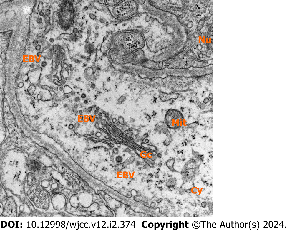
Figure 5 Transmission Electron Microscope revealing the presence of numerous infiltrations of Epstein-Bar virus (80 kV, 40000 ×) within tumor cells.
Nu: nucleus; Cy: Cytoplasm; Gc: Golgi-complex; Mit: Mitochondria; EBV: Epstein-Bar Virus.
- Citation: Kim CS, Choi CH, Yi KS, Kim Y, Lee J, Woo CG, Jeon YH. Absence of enhancement in a lesion does not preclude primary central nervous system T-cell lymphoma: A case report. World J Clin Cases 2024; 12(2): 374-382
- URL: https://www.wjgnet.com/2307-8960/full/v12/i2/374.htm
- DOI: https://dx.doi.org/10.12998/wjcc.v12.i2.374


