Copyright
©The Author(s) 2024.
World J Clin Cases. Jan 16, 2024; 12(2): 374-382
Published online Jan 16, 2024. doi: 10.12998/wjcc.v12.i2.374
Published online Jan 16, 2024. doi: 10.12998/wjcc.v12.i2.374
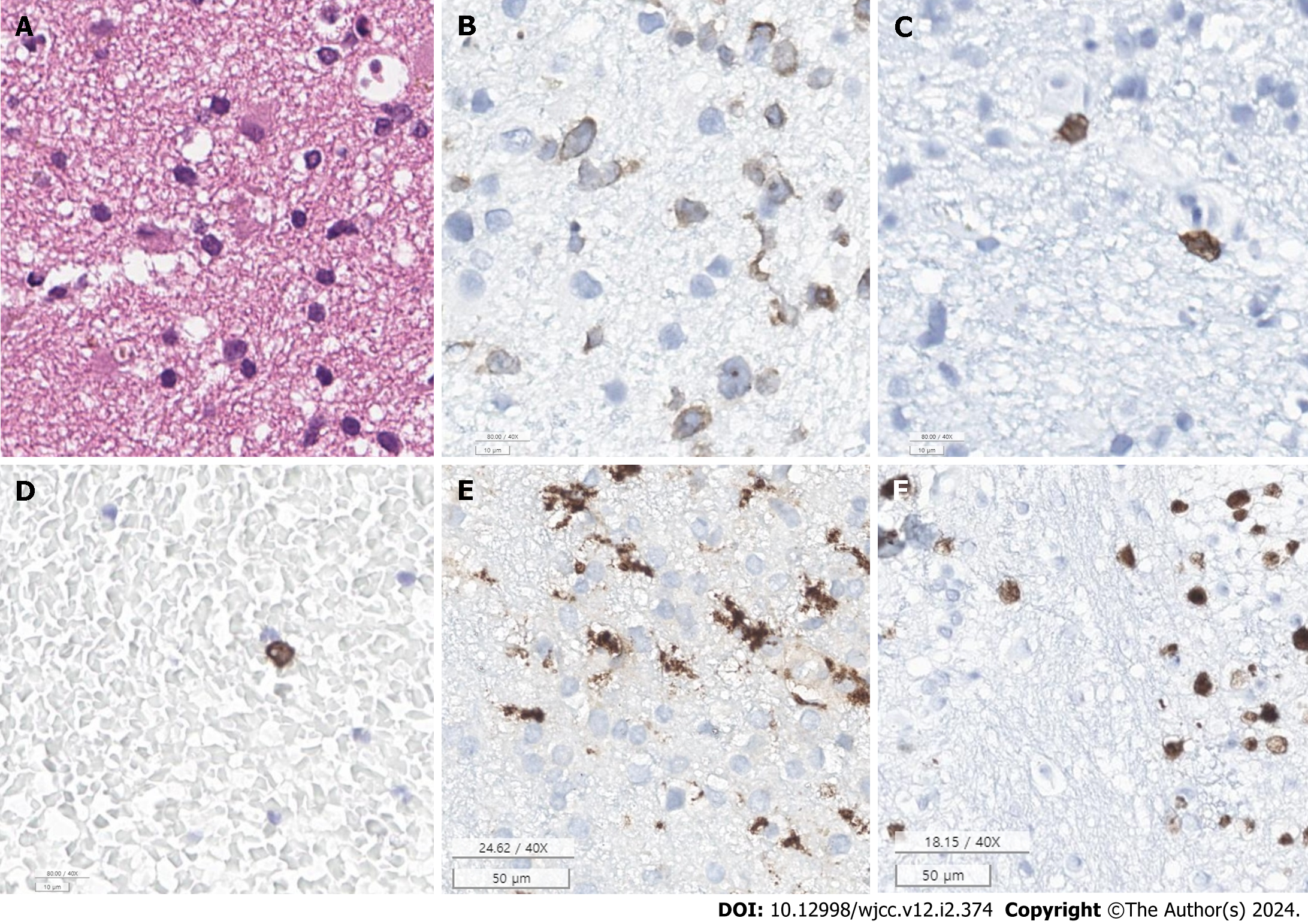
Figure 4 The immunohistochemical staining test results.
A: Hematoxylin & eosin stain (× 40) shows scattered lymphocyte infiltration in the specimen; B: Cluster of differentiation (CD)3 immunostain (× 40); C: CD8 immunostain (× 40), CD3 and CD8 positive lymphocytes suggests T-cell infiltration; D: CD20 immunostain (× 40), CD20 positive cells suggest presence of an immune response, likely towards the neoplastic cells; E: CD68 immunostain (× 40), CD68 positive microglia suggests an ongoing response to an injury or pathology in the brain; F: Antigen Kiel-67 (KI-67) immunostain (× 40), KI-67 index of 40% indicates the presence of high proliferation rate. Overall, these findings suggest a high proliferation neoplastic disorder involving lymphoid cells.
- Citation: Kim CS, Choi CH, Yi KS, Kim Y, Lee J, Woo CG, Jeon YH. Absence of enhancement in a lesion does not preclude primary central nervous system T-cell lymphoma: A case report. World J Clin Cases 2024; 12(2): 374-382
- URL: https://www.wjgnet.com/2307-8960/full/v12/i2/374.htm
- DOI: https://dx.doi.org/10.12998/wjcc.v12.i2.374


