Copyright
©The Author(s) 2024.
World J Clin Cases. Jan 16, 2024; 12(2): 367-373
Published online Jan 16, 2024. doi: 10.12998/wjcc.v12.i2.367
Published online Jan 16, 2024. doi: 10.12998/wjcc.v12.i2.367
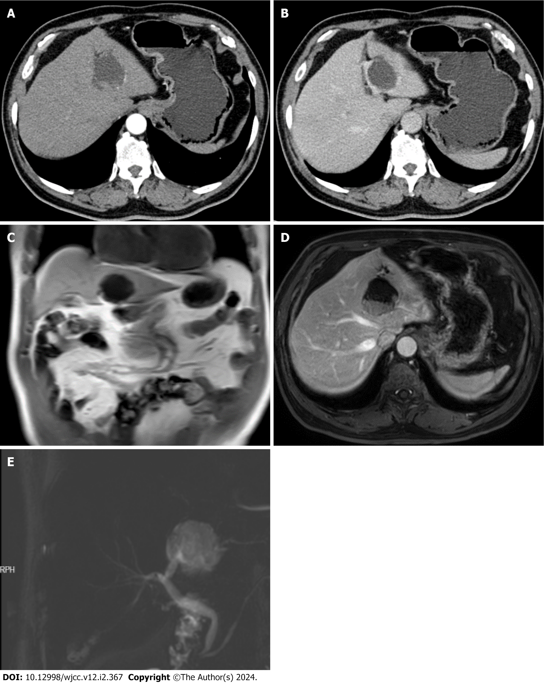
Figure 2 Abdominal enhanced computed tomography and magnetic resonance imaging + magnetic resonance cholangiopan
- Citation: Zhu SZ, Gao ZF, Liu XR, Wang XG, Chen F. Surgically treating a rare and asymptomatic intraductal papillary neoplasm of the bile duct: A case report. World J Clin Cases 2024; 12(2): 367-373
- URL: https://www.wjgnet.com/2307-8960/full/v12/i2/367.htm
- DOI: https://dx.doi.org/10.12998/wjcc.v12.i2.367


