Copyright
©The Author(s) 2024.
World J Clin Cases. Jan 16, 2024; 12(2): 249-255
Published online Jan 16, 2024. doi: 10.12998/wjcc.v12.i2.249
Published online Jan 16, 2024. doi: 10.12998/wjcc.v12.i2.249
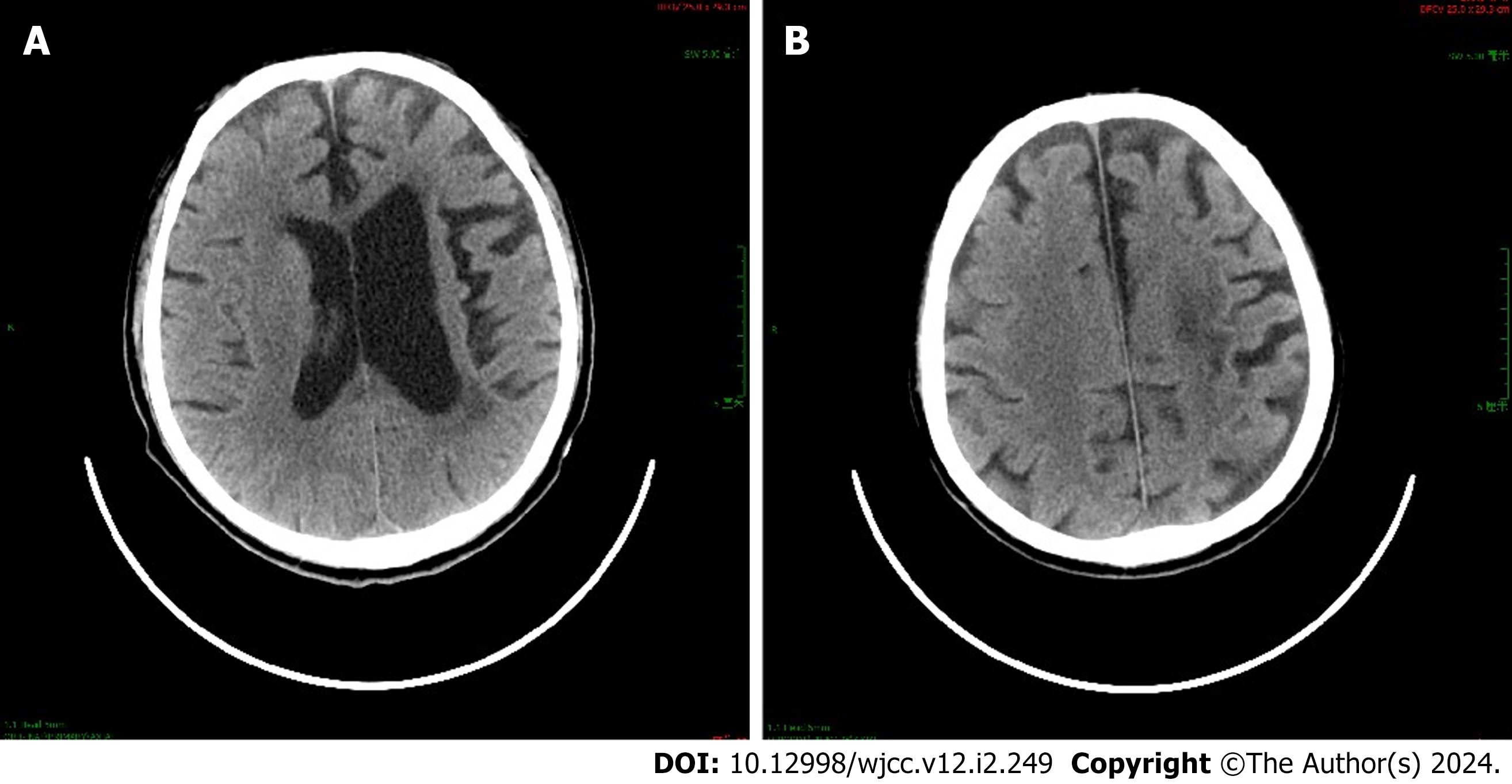
Figure 1 Brain computed tomography of Case 7.
A and B: Brain computed tomography showing multiple patchy, slightly low-density shadows in the left corona radiata (A) and centrum semiovale (B), suggesting old cerebral infarction.
- Citation: Wen LM, Li R, Wang YL, Kong QX, Xia M. Electroencephalogram findings in 10 patients with post-stroke epilepsy: A retrospective study. World J Clin Cases 2024; 12(2): 249-255
- URL: https://www.wjgnet.com/2307-8960/full/v12/i2/249.htm
- DOI: https://dx.doi.org/10.12998/wjcc.v12.i2.249


