Copyright
©The Author(s) 2024.
World J Clin Cases. Jul 6, 2024; 12(19): 3995-4002
Published online Jul 6, 2024. doi: 10.12998/wjcc.v12.i19.3995
Published online Jul 6, 2024. doi: 10.12998/wjcc.v12.i19.3995
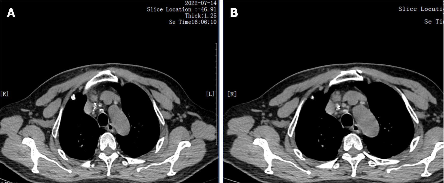
Figure 6 The chest computed tomography mediastinal window.
A: The density of lesions in the posterior segment of the right upper lobe of the mediastinal window is uneven, with calcified shadows visible in some areas; B: Enlargement and calcification of lymph nodes within the mediastinal window.
- Citation: Peng L, Ma R, Li Y, Cheng J. Mycobacterium gordoniasis of the cervical lymph nodes: A case report. World J Clin Cases 2024; 12(19): 3995-4002
- URL: https://www.wjgnet.com/2307-8960/full/v12/i19/3995.htm
- DOI: https://dx.doi.org/10.12998/wjcc.v12.i19.3995


