Copyright
©The Author(s) 2024.
World J Clin Cases. Jul 6, 2024; 12(19): 3995-4002
Published online Jul 6, 2024. doi: 10.12998/wjcc.v12.i19.3995
Published online Jul 6, 2024. doi: 10.12998/wjcc.v12.i19.3995
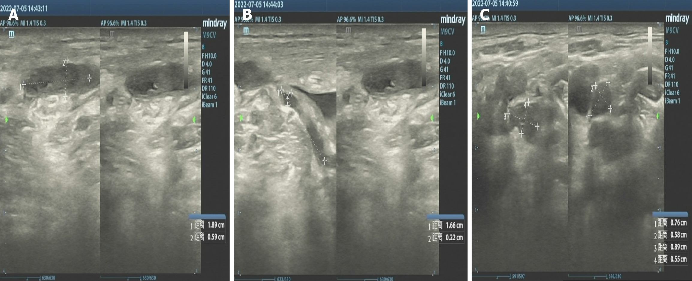
Figure 3 The results of superficial lymph node B-ultrasound.
A: The subcutaneous muscle layer in the lower middle part of the right neck has low echo, with a size of approximately 18.9 mm × 5.9 mm, clear boundaries, and a regular shape; B: The hypoechoic nodule with a width of 2.2 mm and a length of approximately 16.6mm, extending towards the deep left side of the muscular layer; C: The multiple hypoechoic nodule in both neck regions, with clear boundaries and disappearance of cortical and medullary structures, approximately 0.89 mm × 0.55 mm on the left and 0.76 mm × 0.58 mm on the right.
- Citation: Peng L, Ma R, Li Y, Cheng J. Mycobacterium gordoniasis of the cervical lymph nodes: A case report. World J Clin Cases 2024; 12(19): 3995-4002
- URL: https://www.wjgnet.com/2307-8960/full/v12/i19/3995.htm
- DOI: https://dx.doi.org/10.12998/wjcc.v12.i19.3995


