Copyright
©The Author(s) 2024.
World J Clin Cases. Jul 6, 2024; 12(19): 3985-3994
Published online Jul 6, 2024. doi: 10.12998/wjcc.v12.i19.3985
Published online Jul 6, 2024. doi: 10.12998/wjcc.v12.i19.3985
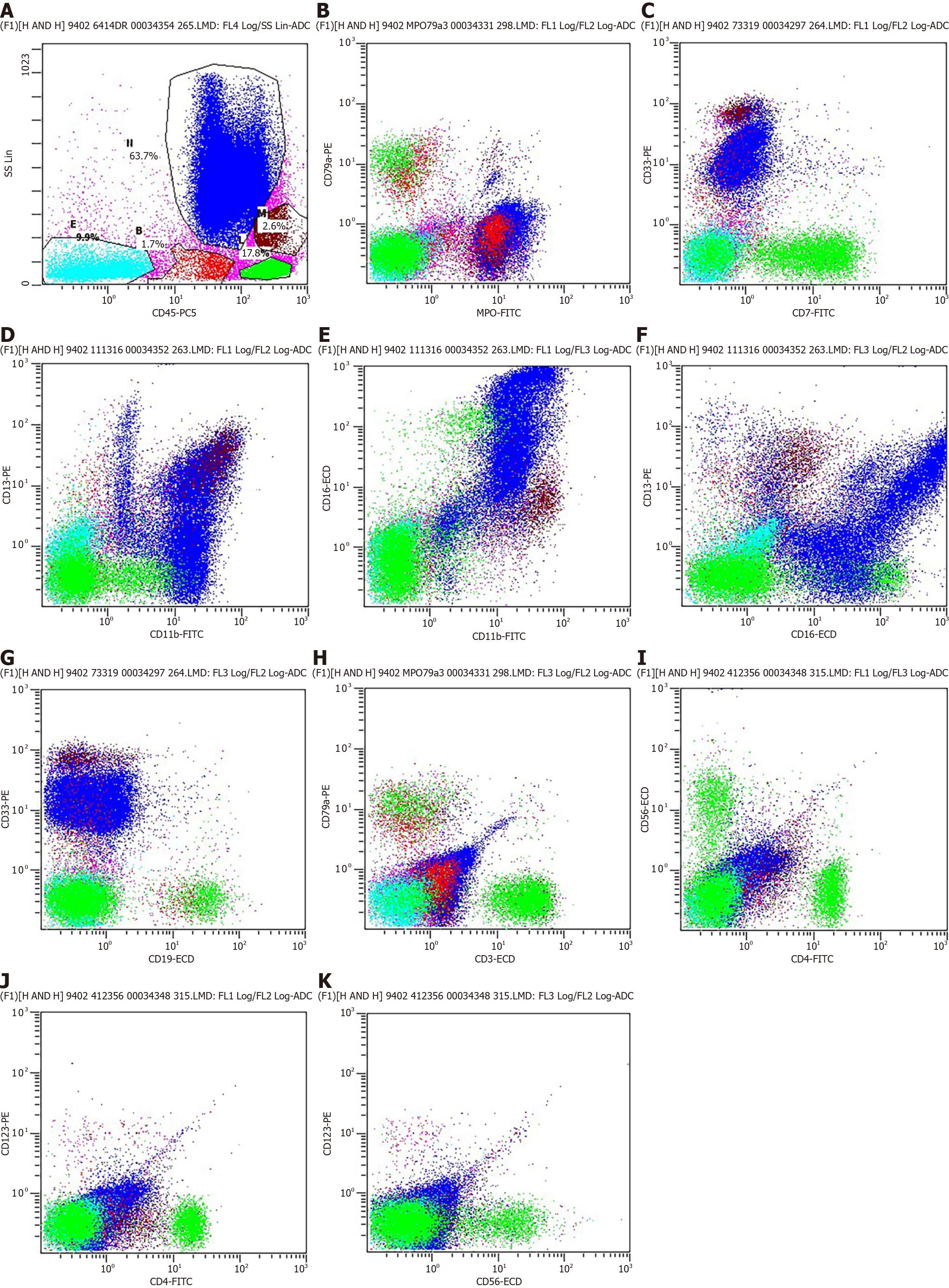
Figure 6 Immunophenotyping using flow cytometry.
A: A 63.7% of the cells were granulocytes, 17.8% of the cells were lymphocytes, 2.6% of the cells were monocytes, 1.7% of the cells were immature cells and 9.9% of the cells were CD45-negative; B-H: Flow cytometry was performed using myeloperoxidase-positive (MPO), CD79a, CD7, CD33, CD11b, CD13, CD16, CD19 and CD3 antibodies. The MPO/CD79a, CD7/CD33, CD11b/CD13, CD11b/CD16, CD16/CD13, CD19/CD33 and CD3/CD79a gate helps distinguish granulocytes morphology. The results showed that granulocytes morphology were generally within normal parameters; I-K: Flow cytometry was performed using CD4, CD56 and CD123 antibodies. The CD4/CD56, CD4/CD123 and CD56/CD123 gate helps distinguish B-cell. No distinct expressing CD4, CD56, and CD123 were observed within the B-cell group in the present study. FITC: Fluorescein isothiocyanate; ECD: Extracellular domain; ADC: Apparent diffusion coefficient.
- Citation: Li SH, Yang CX, Xing XM, Gao XR, Lu ZY, Ji QX. Myeloid sarcoma with maxillary gingival swelling as the initial symptom: A case report and review of literature. World J Clin Cases 2024; 12(19): 3985-3994
- URL: https://www.wjgnet.com/2307-8960/full/v12/i19/3985.htm
- DOI: https://dx.doi.org/10.12998/wjcc.v12.i19.3985


