Copyright
©The Author(s) 2024.
World J Clin Cases. Jul 6, 2024; 12(19): 3985-3994
Published online Jul 6, 2024. doi: 10.12998/wjcc.v12.i19.3985
Published online Jul 6, 2024. doi: 10.12998/wjcc.v12.i19.3985
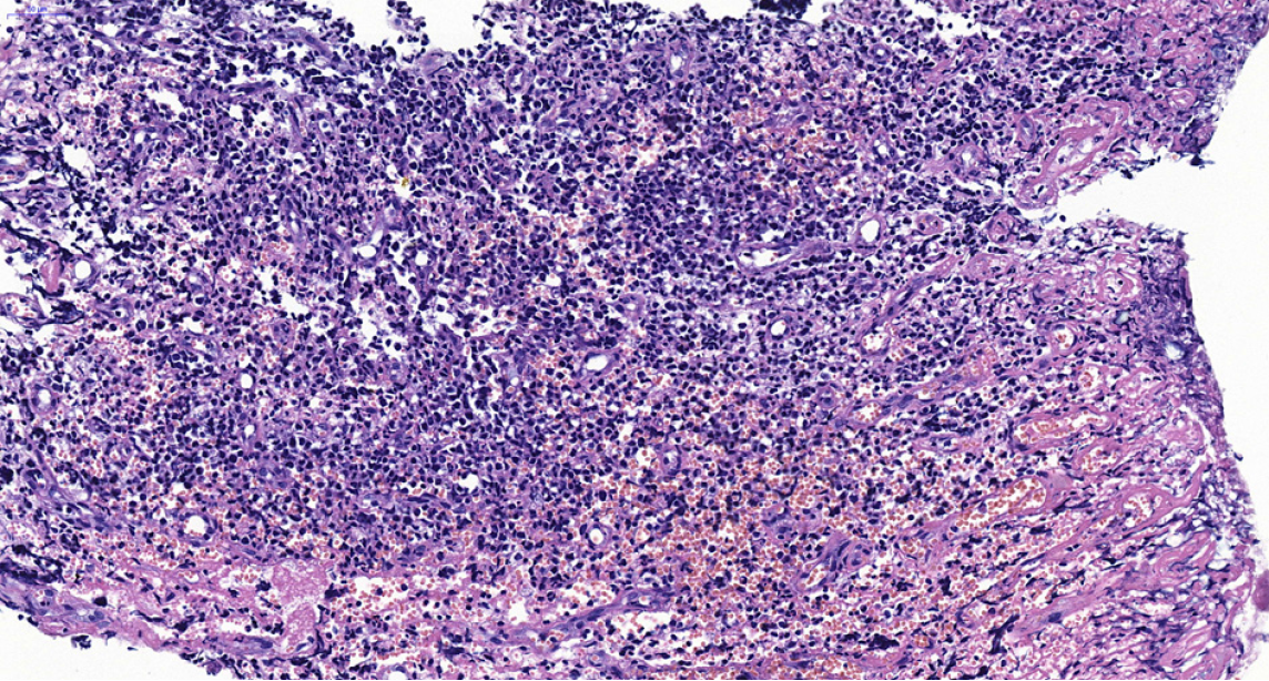
Figure 2 A photomicrograph demonstrated the presence of diffuse neoplastic infiltration in the gingiva, indicating the involvement of abnormal cells that invaded the tissue in a scattered manner.
These neoplastic cells are described as intermediate-sized immature blast-like cells with thin cytoplasm, finely-dispersed chromatin, and rounded or indented nuclei. Magnification, × 300.
- Citation: Li SH, Yang CX, Xing XM, Gao XR, Lu ZY, Ji QX. Myeloid sarcoma with maxillary gingival swelling as the initial symptom: A case report and review of literature. World J Clin Cases 2024; 12(19): 3985-3994
- URL: https://www.wjgnet.com/2307-8960/full/v12/i19/3985.htm
- DOI: https://dx.doi.org/10.12998/wjcc.v12.i19.3985


