Copyright
©The Author(s) 2024.
World J Clin Cases. Jul 6, 2024; 12(19): 3961-3970
Published online Jul 6, 2024. doi: 10.12998/wjcc.v12.i19.3961
Published online Jul 6, 2024. doi: 10.12998/wjcc.v12.i19.3961
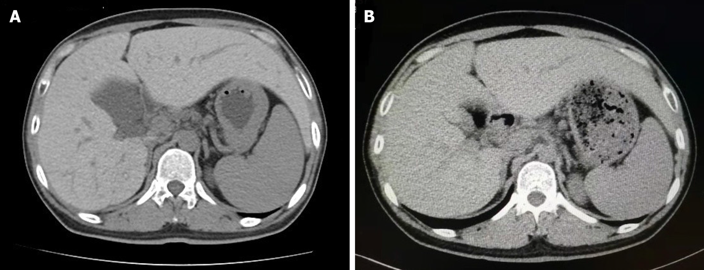
Figure 1 Computed tomography images of the abdomen.
A: Computed tomography (CT) scanning showed that the enlarged liver of the patient clearly appeared as a high-density area because of tissue iron deposition, the CT value of the liver was 104 Hounsfield unit (HU); B: The CT value of the liver decreased to 68 HU after 72 wk of phlebotomy therapy.
- Citation: Xie LD, Kong XM, Shen JX, Wang TL, Ma J, Zhang YF, Chen XP. Novel compound heterozygous mutations in the hemojuvelin gene in a juvenile hemochromatosis patient: A case report. World J Clin Cases 2024; 12(19): 3961-3970
- URL: https://www.wjgnet.com/2307-8960/full/v12/i19/3961.htm
- DOI: https://dx.doi.org/10.12998/wjcc.v12.i19.3961


