Copyright
©The Author(s) 2024.
World J Clin Cases. Jul 6, 2024; 12(19): 3925-3930
Published online Jul 6, 2024. doi: 10.12998/wjcc.v12.i19.3925
Published online Jul 6, 2024. doi: 10.12998/wjcc.v12.i19.3925
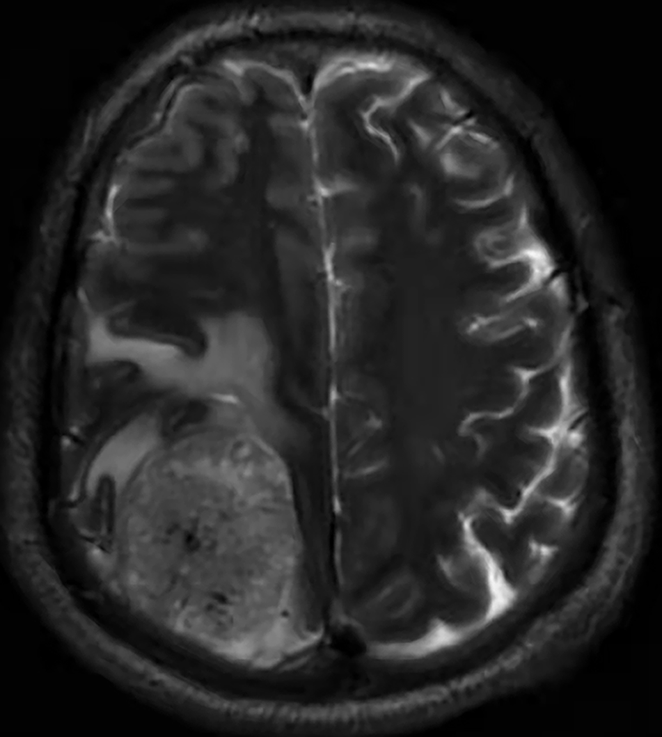
Figure 4 Cranial magnetic resonance imaging on February 16, 2022.
Multiple round mixed signal shadows are detected in the right frontal lobe, temporal lobe, bilateral parieto-occipital lobe, and cerebellar hemisphere. The largest, with a size of approximately 4.6 cm × 4.6 cm × 5.4 cm is located in the right parietal lobe.
- Citation: Xu L, Xu R, Sun J. Anal metastasis in esophageal cancer: A case report. World J Clin Cases 2024; 12(19): 3925-3930
- URL: https://www.wjgnet.com/2307-8960/full/v12/i19/3925.htm
- DOI: https://dx.doi.org/10.12998/wjcc.v12.i19.3925


