Copyright
©The Author(s) 2024.
World J Clin Cases. Jul 6, 2024; 12(19): 3925-3930
Published online Jul 6, 2024. doi: 10.12998/wjcc.v12.i19.3925
Published online Jul 6, 2024. doi: 10.12998/wjcc.v12.i19.3925
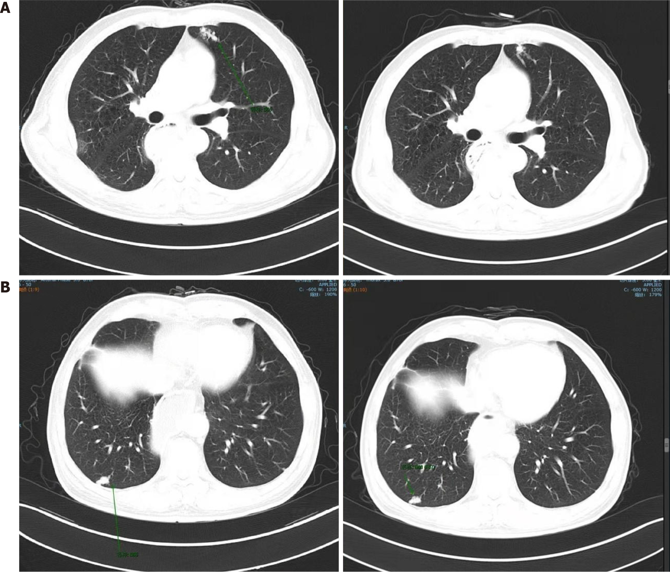
Figure 3 Computed tomography scan of chest and abdomen.
A: Computed tomography (CT) scan of chest and abdomen on September 17, 2019. Soft tissue shadows can be seen next to the spine in the lower lobe of the left lung, along with multiple soft tissue shadows in the lower lobes of both lungs, the largest one being about 1.8 cm in diameter; B: CT scan of the chest and abdomen on December 16, 2019. Multiple patchy and mass soft tissue density shadows are observed in both lungs. The largest one is approximately 3.2 cm × 2.4 cm.
- Citation: Xu L, Xu R, Sun J. Anal metastasis in esophageal cancer: A case report. World J Clin Cases 2024; 12(19): 3925-3930
- URL: https://www.wjgnet.com/2307-8960/full/v12/i19/3925.htm
- DOI: https://dx.doi.org/10.12998/wjcc.v12.i19.3925


