Copyright
©The Author(s) 2024.
World J Clin Cases. Jun 26, 2024; 12(18): 3615-3621
Published online Jun 26, 2024. doi: 10.12998/wjcc.v12.i18.3615
Published online Jun 26, 2024. doi: 10.12998/wjcc.v12.i18.3615
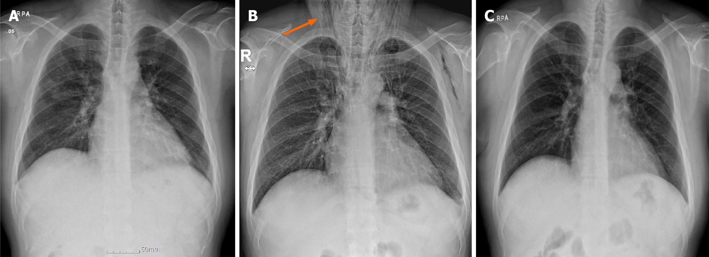
Figure 3 Serial chest radiographs.
A: Chest radiograph taken upon admission showed no abnormalities; B: Chest radiograph obtained after forceful vomiting during bowel preparation showed extensive subcutaneous emphysema in the neck (orange arrow) and widening of the mediastinum, suggestive of pneu
- Citation: Kim G, Lee WH, Kang S, Moon JR, Lee YS, Son JH, Kim NH, Kim JW. Vomiting-induced pharyngeal perforation during bowel preparation for colonoscopy: A case report. World J Clin Cases 2024; 12(18): 3615-3621
- URL: https://www.wjgnet.com/2307-8960/full/v12/i18/3615.htm
- DOI: https://dx.doi.org/10.12998/wjcc.v12.i18.3615


