Copyright
©The Author(s) 2024.
World J Clin Cases. Jun 26, 2024; 12(18): 3589-3595
Published online Jun 26, 2024. doi: 10.12998/wjcc.v12.i18.3589
Published online Jun 26, 2024. doi: 10.12998/wjcc.v12.i18.3589
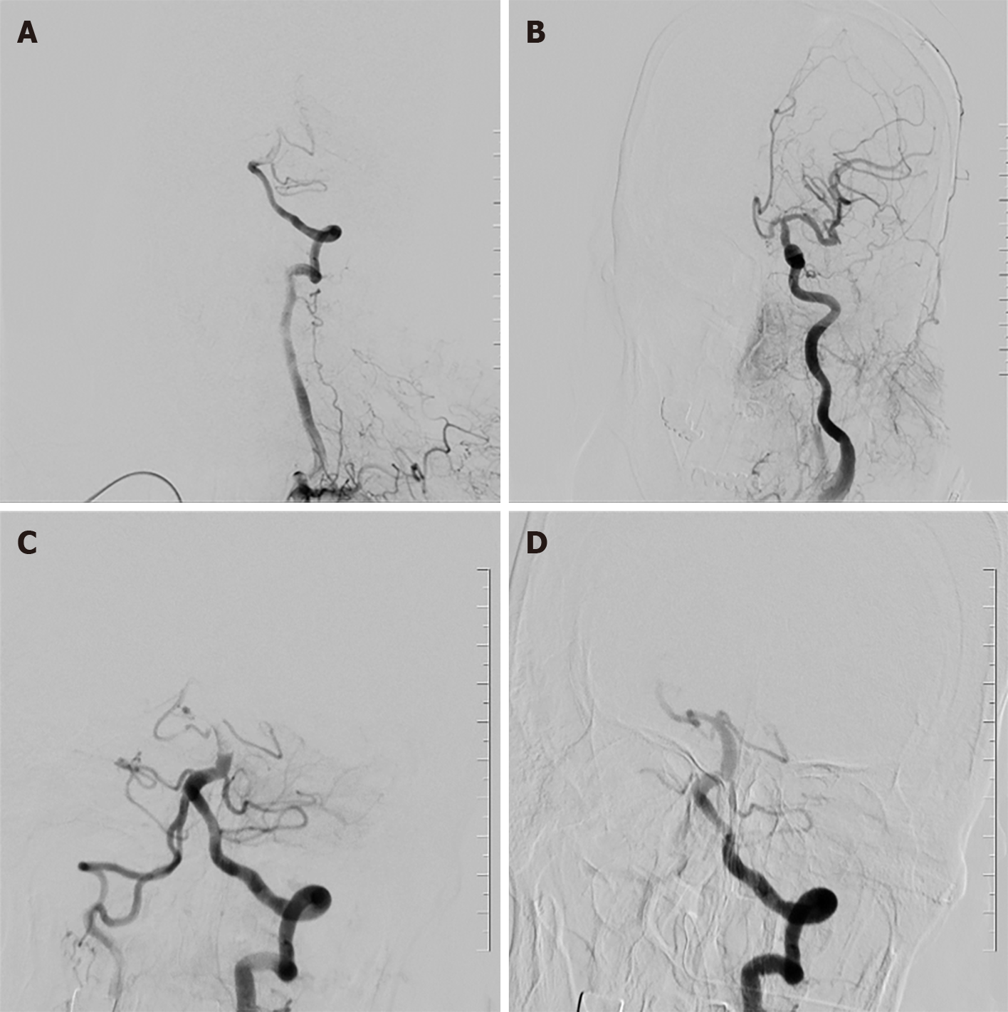
Figure 1 Anterior and posterior thrombograms.
A: The posterior cerebral artery is not developed on the first angiography; B: Left internal carotid artery angiography does not show posterior communication; C: Thrombosis shows basilar artery opening, and positive lateral angiography shows smooth blood flow in the right posterior cerebral artery; D: Occlusion of the posterior proximal segment of the left brain (arrows), and no distant blood vessel is developed.
- Citation: Ding LS, Liang H, Zheng M, Shen M, Li ZJ, Song RP, Chen QL. Extracorporeal membrane oxygenation states basilar artery thrombectomy and left posterior cerebral artery stent thrombectomy: A case report. World J Clin Cases 2024; 12(18): 3589-3595
- URL: https://www.wjgnet.com/2307-8960/full/v12/i18/3589.htm
- DOI: https://dx.doi.org/10.12998/wjcc.v12.i18.3589


