Copyright
©The Author(s) 2024.
World J Clin Cases. Jun 26, 2024; 12(18): 3548-3554
Published online Jun 26, 2024. doi: 10.12998/wjcc.v12.i18.3548
Published online Jun 26, 2024. doi: 10.12998/wjcc.v12.i18.3548
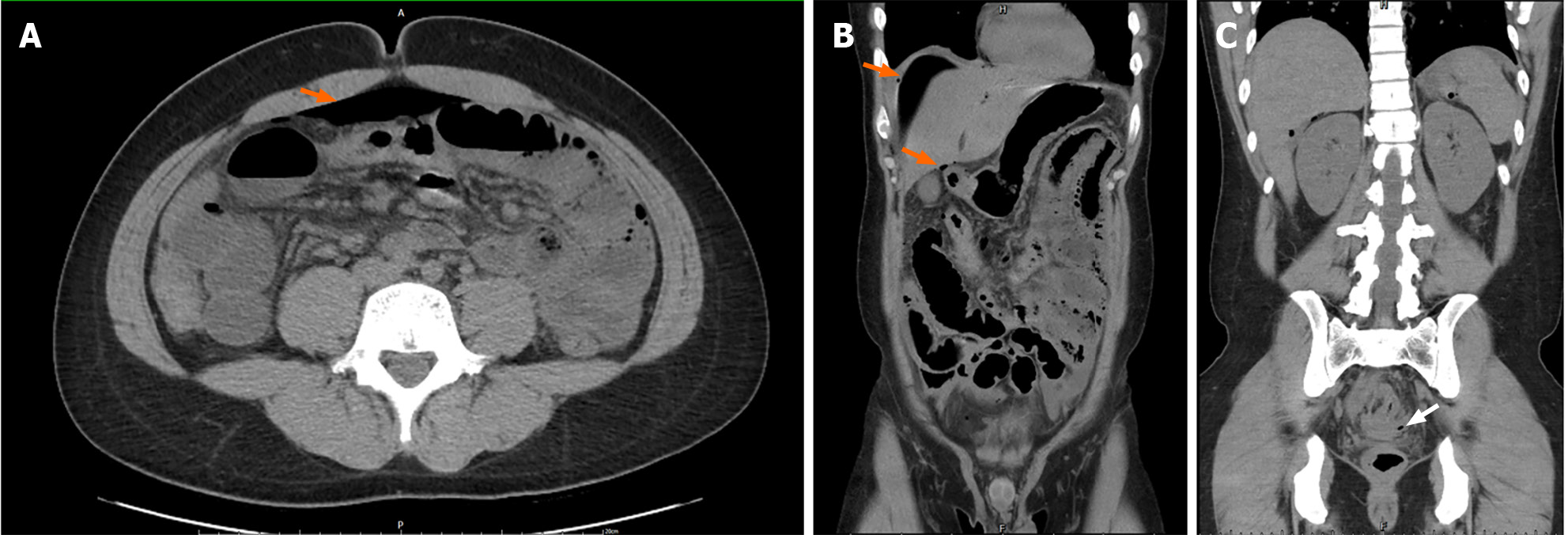
Figure 3 Abdominal computed tomography scan imaging.
A and B: Computed tomography (CT) axial and coronal view showing pneumoperitoneum free air (orange arrow); C: CT coronal view showing fatty stranding around the rectum region (white arrow), but no foreign body was observed.
- Citation: Lin CY, Pu TW. Colon perforation with severe peritonitis caused by erotic toy insertion and treated using vacuum-assisted closure: A case report. World J Clin Cases 2024; 12(18): 3548-3554
- URL: https://www.wjgnet.com/2307-8960/full/v12/i18/3548.htm
- DOI: https://dx.doi.org/10.12998/wjcc.v12.i18.3548


