Copyright
©The Author(s) 2024.
World J Clin Cases. Jun 16, 2024; 12(17): 3253-3258
Published online Jun 16, 2024. doi: 10.12998/wjcc.v12.i17.3253
Published online Jun 16, 2024. doi: 10.12998/wjcc.v12.i17.3253
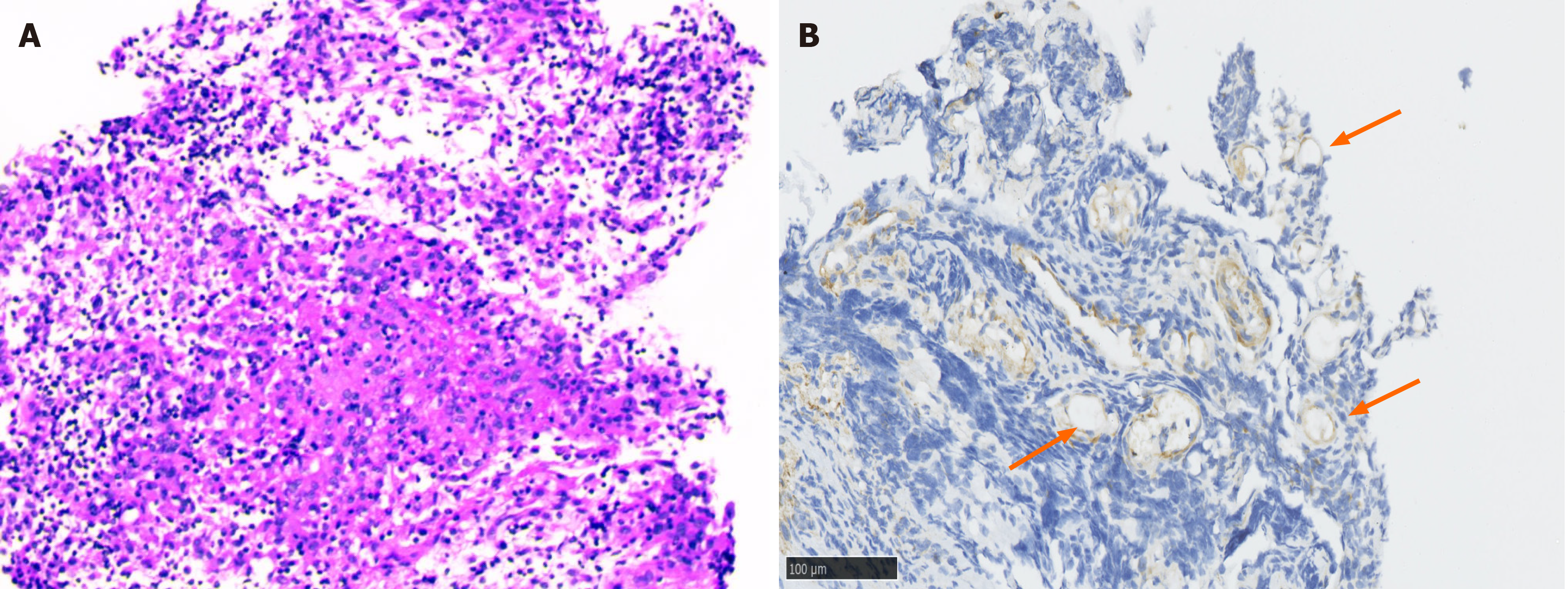
Figure 2 Pathological and immunohistochemical examination results of inflammatory masses.
A: Pathological examination. Lymphocytic infiltration with abundant plasma cells, no eosinophils or microorganisms observed on hematoxylin and eosin staining, original magnification (× 100); B: Immunohistochemical examination. LL-37 positive dilated capillaries (arrows). Brownish-yellow or brown granules indicate positive expression of LL-37.
- Citation: Han XM, Zhou YM, Cen LS. Ocular rosacea without facial erythema involvement manifesting as bilateral multiple recurrent chalazions: A case report. World J Clin Cases 2024; 12(17): 3253-3258
- URL: https://www.wjgnet.com/2307-8960/full/v12/i17/3253.htm
- DOI: https://dx.doi.org/10.12998/wjcc.v12.i17.3253


